Tag Archives: Amygdala

A Review on Brain’s Health

As much as there have been many suggestions and studies on how to cope with the pandemic using multi-modal means, such as apps, vitamins or physical exercise, or even new hobbies and food adventures. And that is on the upside of things. Coping skills when the cards are stacked against your favor hyper focuses on survival from moment to moment when the brain is overloaded and tasked with overchoice.

Multi-million-dollar industries have been built on people’s pursuits on the fountain of youth and keeping the brain young or at least to match the youthful vigor of how the body should feel. Emotionally despite our wanting for vigor, we are brought back to realization by our brain’s level of resources or how to retrieve it as the amygdala automatically pushes past the suggestion of pain or difficulty when basic needs are of the essence.

Caring for the brain’s emotions are interdependent on the human capacity for social cognition when reading body language and expressions. And in the stressful pandemic times, adaptive social behavior and mental well-being also require inferring the absence of emotional content. Of relevance during exceptional events such as the coronavirus disease 2019 (COVID-19) pandemic involving social distancing and isolation as well as higher levels of anxiety but also, requiring intact social communication and empathy. (Sokolove et al, 2020)

When one is required to maintain social distance and/or uses PPE while interacting with another, the interplay between the amygdala and insula is elevated as it contributes substantially to the processing of social signals that convey emotionally neutral information, underwriting a fundamental yet underestimated component of adaptive social cognition and behavior. The absence of stimulatory emotion presents a difficulty of processing in the bond communication between the amygdala and insula with the ability to infer the
absence of emotion in body language, including with the processing within the amygdala and insula have been associated with emotion perception. Ultimately, when caring for the brain’s health scanning other people’s words and delivery are what is needed for homeostasis.

So, the brain begins searching for alternates in reading and writing ’emotion’ in times of crises to maintain itself. Depending on the coping mechanisms embedded within the connections, physical and/or psychological experience that binds with an external event, this one being the lack of neurotypical emotion perception could potentially ripple into pre-existing trauma. There are growing indications that these trauma-exposed individuals show
neural alterations, even in the absence of clinical symptoms and tasks requiring the regulation of emotions and impulses seem to be particularly affected. Sabrina Golde et.al

studied 25 women (mean age 31.5 ± 9.7 years) who had suffered severe early life trauma but who did not have a history of or current psychiatric disorder, with 25 age- and education-matched trauma-naïve women by utilizing fMRI study investigated response inhibition of emotional faces before and after psychosocial stress situations in 2019. In both groups, nontraumatic stress significantly impaired response inhibition of fearful facial expressions but did not affect inhibition of happy or neutral stimuli. When inhibiting responses to fearful faces under stress, women who had experienced severe and multiple traumatic events before the age of 18 demonstrated a decreased brain signal in the left inferior frontal gyrus in the frontal lobe, but a marginally increased signal in the right anterior insula (between the frontal and temporal lobes) compared to trauma-naïve control subjects.

They also discovered that in trauma-naïve control subjects only, the inferior frontal gyrus activation was linked to a lower rate of stress-induced false alarms, whereas in women who had experienced severe and multiple traumatic events before the age of 18, insula activation was linked to a higher number of false alarms.

Of course, that begs the question, would brain health be different between a male brain in comparison to the female brain? Are brains not simply just as generic as other parts of the body such as the skeletal and muscular systems? Would the same study above if applied to men render equitable results as the women have displayed? Perhaps, as brain function is dependent on synapses and stimuli response. However as unique as every individual is, the responses can and will be varied according to the emotional pairing, regardless of assigned biological sex.

After all neuroplasticity happens to people, mostly in the background, positive or negative. The learning experience is constantly evolving, including in what is automatic such as driving, to the more complex tasks such as learning a new skill or trade. Yet in the viewpoint from a healthy brain, neuroplasticity would support neural pruning at every stage of life: keeping only the connections that are meaningful and consistently used for an end goal while allowing brain real estate to grow for additional new experiences to bind. If, however the exposure is constantly negative neuroplasticity such as death, destruction, or violence, the brain’s stress levels are increased thus decreasing overall expanded connections and brain weight.

Sandra Racionero-Plaza, et al., found in their research article, “Architects of Their Own Brain.” Social Impact of an Intervention Study for the Prevention of Gender-Based Violence in Adolescence sought to review the social impact of psychology in the field of teen gender violence from participants in the MEMO4LOVE project comprised 126 adolescents attending three different high schools in Barcelona (Spain). The aim was to report on the potential social impact achieved by an intervention study consisting of seven interventions on the preventive socialization of gender violence, with specific interventions focused on seven different topics. Part of the intervention were dimensions of the influence of the dominant coercive discourse in adolescents’ sexual-affective life, accompanied by egalitarian dialogue with the adolescents on how to break free from such discourse and thus avoid violence in sporadic or stable sexual-affective relationships. Below is what they found:

1) The adolescents who took part in these interventions reported developing critical consciousness regarding the presence of the dominant coercive discourse in society and in their lives. The greater the transformation of peer interactions, the greater the prevention of gender-based violence victimization.

2) It is necessary that intervention strategies in education provide adolescents with tools that enable them to be more critical about violence in society. Interventions provided them with cognitive tools to better analyze their own and others’ sexual-affective thinking, emotions, and behaviors in ways that favor the rejection of violent males and violent sexual-affective relationships

3) Change in preferences and attraction, with greater rejection of violent men and enhanced attraction toward egalitarian masculinities. It is significant to be aware of the dominant coercive discourse, but what is even more fundamental is to change the response of attraction toward men with violent attitudes and behaviors; that is what will make a difference in terms of preventing gender-based violence victimization.

Society therefore continues to impact the neuroplasticity quality with our degrees of interaction with it. Aside from gender and age groups, it is of significance to mention that brain health across races also have complex layers affecting neuroplasticity. In the research article, Insights From African American Older Adults on Brain Health Research Engagement: “Need to See the Need” Soshana Bardach, et al., posited that African Americans (AAs) have an elevated risk of developing dementia, yet are underrepresented in clinical research. In order for the 21 AAs aged 35-86 years to agree to be part of the study, the researchers had to ensure that the facilitators were AA from their community, laying out clearly and plainly the methodology that would be used to gather data which is called Photovoice, and to be transparent about the reasons behind the data gathering.

The use of Photovoice in data collection is a community-based, participatory-action research methodology, centered on the premise that sharing perceptions and experiences through photography and discussion leads to an awareness and understanding of relevant issues (Harley et al, 2015) It was through this method that the discussion of brain health research among AAs highlighted several barriers to participation and corresponding strategies for addressing the identified barriers and encouraging engagement. The barriers and solutions were in the areas of (a) mistrust stemming from historical mistreatment and reinforced by contemporary injustices, (b) avoidance and fear of acknowledging problems such as being afraid of what they may find out or not wanting to know as barriers to seeking a diagnosis, and (c) seeing the risks of research, but not the need as they anticipate potential unpleasant aspects of research participation were more tangible than the benefits.

This then brings us to consider that brain health is not only a result of positive neuroplasticity but a contiguous history of sustainable emotions stemming from society and communities. The deterioration of the brain affected by genetics is also sped up by neurochemicals that inflame the stress response on a consistent basis thus making recovery to a biologically sound baseline unattainable. Anti-declining the brain, simply put, is to bring it to a social balance. An immediate way is to encourage pro-social behaviors between humans and/or animal species. Pro-social behaviors—actions that help others, often without regard to whether they directly benefit us—are vital for social cohesion. (Lockwood, et al, 2020) The idea is that to do acts that benefit other people, we first must learn what actions in the world will lead to something rewarding and positive happening to them, all processed in the anterior cingulate cortex of the brain. When we perform an action, and it results in a reward for another person, it is easy to imagine ourselves in their shoes and vicariously experience what it would be like to get a similar reward ourselves.

If there are limited opportunities for vicarious learning or pro-social behaviors, such as when in a global crises, how can balance be achieved? Ironically, Polyphenol-Rich Interventions are in order to influence the sensitivity of natural neurotransmitters and adjust levels naturally. Polyphenol compounds, such as flavonoids, phenolic acids and tannins, are found in varying concentrations in a range of plant-based food sources (e.g., legumes, fruit, vegetables, herbal extracts, spices, coffee, tea and cocoa), and their effects on human health have drawn considerable attention. (Ammar, et al 2020). It is the self-socializing method by virtue of self-care that supports choices of the quality of brain health. At this particular juncture, the use of the human amygdala is its response to other emotionally significant stimuli beyond threat, including positive or reward information, but also novelty and non-emotional salient stimuli with personal impact or goal-related significance. This diversity of response patterns has led to recent theoretical accounts proposing that the amygdala may actually encode the “relevance” of events, which is determined by the goals, needs, or values of the individual, in a context-dependent manner. (Guex et al, 2020) This account accords with psychological theories, such as the component process model of emotional appraisal as would in patterns of self-socialized self-care.

The anterior cingulate cortex and the amygdala would need support when communicating emotions-bound signals to each other. It is with the subcortical structures of the basal ganglia (BG) and the cerebellum that support multiple domains including affective processes such as emotion recognition, subjective feeling elicitation and reward valuation. By regulating
cortical oscillations to guide learning and strengthening rewarded behaviors or thought patterns to achieve a desired goal state, these regions can shape the way an individual processes emotional stimuli. (Pierce J, et al 2020) In essence, no matter how much objective data could be presented to standardize the ideal brain health for a given time frame, developmental stage, race or gender, the conclusion will rest upon every individual’s experience or norm of how their brains are in status quo or if they are on polar opposites. Whatever the case may be, brain health is essential for quality of life.

Learning’s Many Costs, First Quarter 2015 Edition

Flipping channels, Dr. Phil comes onto the main screen and has a mother and daughter in opposite angles to him. The problems seemed to be typical psychological, reality-show formula: mom is overprotective of her teenage daughter, teenage daughter ‘rebels’ and begins a long distance relationship with a man 10 years her senior (who also happens to be a drug dealer still serving time), and the conflicts between them become unbearable. Dr. Phil in his infinite wisdom brought out his objectivity to the mother, and then to the daughter. It is however the explanation to the daughter that resonates with her: “Now remember, it is the Neocortex that develops last in the brain, and that means right now, you’re not able yet to have complete insight to all of the consequences of your actions. This is why you would need guidance with some of your decisions, including starting a relationship with a currently jailed drug dealer.”

Most of us have a familiarity with family drama played out on television.  Whether the purpose is to entertain or to educate, the family’s dysfunctional dynamic from one to the next seem to be rooted deep into social, educational and economic factors. How then is the brain development affected? Is Dr. Phil’s approach by combining the brain science with family dynamics warranted?

money-house

In the early online edition of the journal Nature Neuroscience (March 30, 2015), investigators from nine universities across the country reports correlative links between family income and brain structure. Relationships between the brain and family income were strongest in the lowest end of the economic range, thus suggesting that interventional policies aimed at these children may have the largest societal impact. The study, led by researchers at The Saban Research Institute of Children’s Hospital Los Angeles and Columbia University Medical Center.

In the largest study of its kind to date, the researchers looked at 1,099 typically developing individuals between the ages of 3 and 20 years as part of the multi-site Pediatric Imaging, Neurocognition and Genetics (PING) study. Associations between socioeconomic factors (including parent education and family income) and measurements of surface area of the brain were drawn from demographic and developmental history questionnaires, as well as high-resolution brain MRIs. Statistics (controlled for education, age and genetic ancestry)  showed that income was nonlinearly associated with brain surface area, and that income was more strongly associated with the brain than was parental educational attainment.

First author Kimberly G. Noble, MD, PhD says, “Specifically, among children from the lowest-income families, small differences in income were associated with relatively large differences in surface area in a number of regions of the brain associated with skills important for academic success. ” Dr. Noble is an assistant professor of pediatrics and director of the Neurocognition, Early Experience and Development (NEED) Lab of Columbia University Medical Center. She is also an associate professor of Neuroscience and Education at Teachers College, Columbia University.

Conversely, among children from higher-income families, incremental increases in income level were associated with much smaller differences in surface area. Higher income was also associated with better performance in certain cognitive skills; cognitive differences that could be accounted for by greater brain surface area.

“While in no way implying that a child’s socioeconomic circumstances lead to immutable changes in brain development or cognition, our data suggest that wider access to resources likely afforded by the more affluent may lead to differences in a child’s brain structure,” said Elizabeth Sowell, PhD, director of the Developmental Cognitive Neuroimaging Laboratory, part of the Institute for the Developing Mind at CHLA.  “Family income is linked to factors such as nutrition, health care, schools, play areas and, sometimes, air quality,” added Dr. Sowell, indicating that everything going on in the environment shapes the developing brain. “Future research may address the question of whether changing a child’s environment — for instance, through social policies aimed at reducing family poverty — could change the trajectory of brain development and cognition for the better.”

From the socio-economic factors, we take a look at socio-psychological factors, some of which affect our ability to create meaningful connections. A recent study from the University of Georgia shows differences in brain structure according to how trusting people are of others.

The team of researchers used two measures to determine the trust levels of 82 study participants. The participants filled out TRUSTING CHILDa self-reported questionnaire about their tendency to trust others. They also were shown pictures of faces with neutral facial expressions, and were asked to evaluate how trustworthy they found each person in the picture. This gave researchers a metric, on a spectrum, of how trusting each participant was of others.

Researchers then took MRI scans of the participants’ brains to determine how brain structure is associated with the tendency to be more trusting of others. What researchers found were differences in two areas of the brain.

“The most important finding was that the grey matter volume was greater in the ventral medial prefrontal cortex, which is the brain region that serves to evaluate social rewards, in people that tended to be more trusting of others,” said the study’s lead author Brian Haas, an assistant professor in the department of psychology.

“Another finding that we observed was for a brain region called the amygdala. The volume of this area of the brain, which codes for emotional saliency, was greater in those that were both most trusting and least trusting of others. If something is emotionally important to us, the amygdala helps us code and remember it.”

The long term hope for the research may have implications for future treatments of psychological conditions such as autism. Future studies may focus on how, and if, trust can be improved and whether the brain is malleable according to the type of communication someone has with another. “There are conditions, like autism, that are characterized by deficits in being able to process the world socially, one of which is the ability to trust people,” Dr. Haas said. “Here we have converging evidence that these brain regions are important for trust; and if we can understand how these differences relate to specific social processes, then we may be able to develop more targeted treatment techniques for people who have deficits in social cognition.”

So what can we do as a community with fragile families who have young children in such a fast-paced, competitive, and digitally plugged world? Begin the developmental awareness young for a firmer foundation with research-backed programs.

Supporting this is new research from UNC’s Frank Porter Graham Child Development Institute (FPG) revealing high-quality early education is especially advantageous for children when they start younger and continue longer. Not only does more high-quality early education significantly boost the language skills of children from low-income families, children whose first language is not English benefit even more.

“These findings show that more high-quality early education and care can narrow the achievement gap before children reach kindergarten,” said Noreen M. Yazejian, principal investigator of FPG’s Educare Learning Network Implementation Study. “Children from low-income families can improve their standing relative to their middle class peers.”

Ms. Yazejian said previous research has shown language skills are most malleable for children before age 4, which in large part explains high-quality early education’s powerful effects. Her study examined children’s receptive language skills–the ability to hear and understand words–because these particular skills are an excellent predictor of later academic success.

According to Yazejian, Educare classrooms offered the chance to study children enrolled in high-quality early education and care from the earliest ages. Educare is an enhanced Early Head Start and Head Start program for low-income, high-needs children from 6 weeks old until entry into kindergarten. The model has been replicated in 20 schools nationwide over the last 15 years.

“Educare’s comprehensive approach to early childhood education aims to level the playing field for children living in poverty,” said Portia Kennel, executive director of the Educare Learning Network. “This new study confirms that we need to include the earliest years of life as part of our nation’s education system. Quality early education prepares vulnerable children for success by preventing the achievement gap that appears long before kindergarten.”

Many people traditionally have viewed early care for infants primarily as a support for mothers who want to work and not as an essential component of early schooling. However, findings from the FPG study add to a growing body of research revealing better outcomes for children from low-income families who start high-quality education earlier and stay in it longer.

Earlier research has shown the English language skills that dual-language learners develop prior to kindergarten can predict educational achievement through eighth grade, but keeping skills in the home language also is beneficial. Home language skills are related to long-term social, emotional, cognitive, and academic outcomes.

“Most dual-language learners in this study were in classrooms where English was the primary instructional language but in which one staff member could use their home language as needed to support learning,” Ms. Yazejian explained. “It’s not surprising our findings show they quickly acquired skills in English. That’s why it’s reassuring that our study found that the acquisition of English language skills in Educare classrooms does not come at the expense of Spanish skills.”

The number of young children who speak a language other than English at home is growing, and this study contributes valuable new information to the field. “It’s encouraging to see that dual-language learners are making strides that form the critical foundation for later learning,” according to Ms. Kennel.

Ms.Yazejian encourages the thinking that more than one year of high quality early care and education brings greater benefits for children. “The differences we found in this study, extrapolated to thousands of children–especially dual-language learners–could add up to lasting effects and lower public education costs.”

cropped_77388_Royalty_Free_RF_Clipart_Illustration_Of_A_Diverse_Group_Of_Children_Spelling_The_Word_Preschool1

Is it Negative Behavior or ADHD Sensory Overload? An Educator’s Quick Reference

How many times have students been pigeon-holed into the category of displaying bad or negative behavior when opposing class work or during transitions from a state of play or break back to the classroom and vice versa?

When the body appears like this during an overt meltdown:

What May Look Like This May Actually Not Be...
What May Look Like This May Actually Not Be…

The Brain Actually looks like this:

The Amygdala and Hypothalamus Fired Up in Fight or Flight State

The Emotional Brain that is highlighted are two specific parts of the limbic system, the amygdala and the hypothalamus. The amygdala controls the brain’s ability to coordinate many responses to emotional stimuli, including endocrine, autonomic, and behavioral responses. Stress, anxiety, and fear are primary stimuli that produce responses. Mediation by the amygdala allows control among the stimuli.

The hypothalamus plays a significant role in the endocrine system and are effected by the amygdala. It is responsible for maintaining your body’s internal balance, which is known as homeostasis. This includes the  heart rate, blood pressure, fluid and electrolyte balance, appetite, sleep cycles and is the key connector between the endocrine system (glands and hormones) and the nervous system.

Now we are painting this picture of the brain developing at a functionally optimal manner; without aberrations from either genetic means or environmental factors. However, when faced with students who have underlying imaging differences in brain imaging due to the said factors and manifest a type of negative behavior that can easily be mistaken and categorized as a regular tantrum, the subtle elevations in amygdala and hypothalamic responses are now pushed to abnormally erratic levels in these brains.

For example, take the Attention Deficit Hyperactivity Brain in comparison to the Normal Brain:

We see clearly that the shape alone of the cerebrum of the ADHD brain is not elongated or similar to a normal brain’s saddle

Imaging of the Normal Brain in Contrast to the ADHD brain

type shape. It is oblong and with heavy concentration on temporal and occipital real estate versus the butterfly formation of the normal brain. What is also fascinating is the corpus callosum (where part of the amygdala and hypothalamus are housed) is lighter in the ADHD brain. What that means is that there is no clear path of communication between both hemispheres as compared to that of a normal brain. The blues indicate calm sections of the brains and the greens are considered to be the brain in an even keeled state, balanced and not in fight-flight mode.

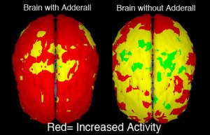
Here’s also an image of a person with and without ADHD medication:

Brain Chemical Responses with Adderall Versus Without Adderall

With Adderall, the brain is utilized in full functional capacity, the chemical connections between neurotransmitters is efficient and there are little if any underutilized processing areas. When Adderall is wearing off, the results are unimaginable: the only sections  of the brain that have any residual function left are the orbitofrontal area of the Pre Frontal Cortex (responsible for sensory integration and some decision making), and spotty areas across the 4 lobes. What is fascinating to mention here is the loss of Adderall effects are from back to front of the cerebrum.

These images provide a very clear picture of the typical versus atypical brain, especially the differences between one with ADHD and one without.   If ony it were that easy as a classroom teacher to distinguish a student with ADHD from a student with  sensory overload.  The list below is not as ‘yellow’ and ‘red’ as the brains above, but hopefully it will provide clarity and a concrete direction for you to take in order to best meet the needs of your students.

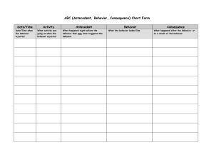
First, it crucial to note that boys and girls with ADHD display different symptoms; therefore, they are distinguished below.  Second, students with meltdowns as a result of negative behavior, will most likely present with similar symptoms; therefore, it is an undertaking for teachers to take quantitative data on the targeted behaviors. Forms like the one below:

TRUE ABC Chart For Objective DATA Collection
TRUE ABC Chart For Objective 5 Session DATA Collection (click for printable image)

BOYS

  • Fidgety while sitting
  • Talk nonstop
  • Constant motion, may include touching items in their path
  • Difficulty sitting still
  • extreme impatience
  • Always “bored”
  • Lack verbal filter
    Sensory Overload or Negative Behavior?
  • Interrupt others’

GIRLS

  • Spacey
  • Unfocused
  • Inattentive
  • Trouble with organization
  • Forget directions
  • Forget or incomplete homework
  • Lose or misplace papers, books, personal belongings
  • Much Less Likely
    • hyperactive
    • impulsive

For students with ADHD, these symptoms as well as sensory overload meltdowns will be manifested consistently throughout the day across environments, unless the student is highly engaged in a preferred activity. Students presenting with negative behaviors will have meltdowns at specific yet intermittent periods of the day or throughout the day as will be shown in the ABC Chart above. For example, when the medication is wearing off, one may see a spike in ADHD symptoms in any combination. Once you can answer when, where, how long and make valid hypotheses as to why students are displaying the behaviors below, you should be able to have a pretty strong understanding as to whether your student is having a meltdown because of learned negative behaviors or as a result of having an ADHD brain on sensory overload.