Category Archives: Brain Bits

Fragile Flexibility (EF Skills Series)

In young children, the schema of their quality world usually revolves around a caregiver or a person who they consider as important in the development of their identity. Their interests initially mimic from imagining that they are versions of the adults they are surrounded by until they are exposed to wider environments, peers, language, media, and then a wholistic interest database emerges from the conglomeration and exposure.

It also makes sense that the younger the child, the more questions they ask. Rarely would you find a child between the ages of 3-5 years come into contact with adults who have set values or biases of themselves concerning what’s ‘right’ or ‘wrong’ around the way they perceive the world. This type of mental flexibility, of mixing and matching new, or new-old ideas in their youth is also directly proportional to the physical activity that they engage in. The pushing of limits of their physical capacities around places that are close to the natural world like garden parks, or places that have been landscaped for the socialization of little people like urban parks are indicators of their levels of mindful curiosity.

It certainly becomes less correlational when children become older, the degree or type of questions with physical movement. The overt questions may turn into musings and conformity is usually expected when school-age commences. Physical movement is timed if the child is not involved in organized sports or games. Just the same, however, the degree of flexibility in these minds depends on the environment they take in, and the imagination that is left from viewing the world from their youth. Martinez and Riba’s 2021 study, Cognitive Flexibility in Schoolchild Through the Graphic Representation of Movement postulates that Neuroconstructivism is the progressive complexity of mental representation over the course of cognitive development and the role of the graphic representation of movement in the transformation of mental schemas, cognitive flexibility, and representational complexity.

They also discuss that In this differential trajectory, mental representation is a key element for cognitive development and for understanding the emergence of child drawing, and changes thereof, as a graphic representation of internalized models of reality (Sirois et al., 2008). A child’s drawing is the first marker that enables the study of mental representation as an external manifestation of internalized reality, by showing what is known about it.

Moreover, events are naturally more attractive than objects, and their foremost feature is their movement. Therefore, part of the content of the first mental representations turns around the identity of events, objects, and people, and their movement and position, which forms the basis of the dynamic representations produced. The first external representative manifestation is the child’s scribble, in which the action of the drawing already contains expressive and representational meanings relating to shapes, movements, and emotions (Quaglia et al., 2015), even if there is no real figure that relates to a meaningful movement for representational purpose.

Such cognitive flexibility is what drives competition in a crowd. The narratives that may have been handed down from authority figures that were used to set ‘safety’ limits, such as limiting or eliminating outdoor time due to the location of where the child resides, or in this recent case the pandemic, inadvertently have pared down the curiosity factor toward the external influences. Subsitutions by devices and programs on the web were meant to digitize the parallel experience of the world beyond the home, however, without the multisensorial inundation of an experience, the ideas being written are almost dream-like. They may be able to describe a forest of trees in a contextual litany of facts, but ask them about the experience and then they are puzzled.

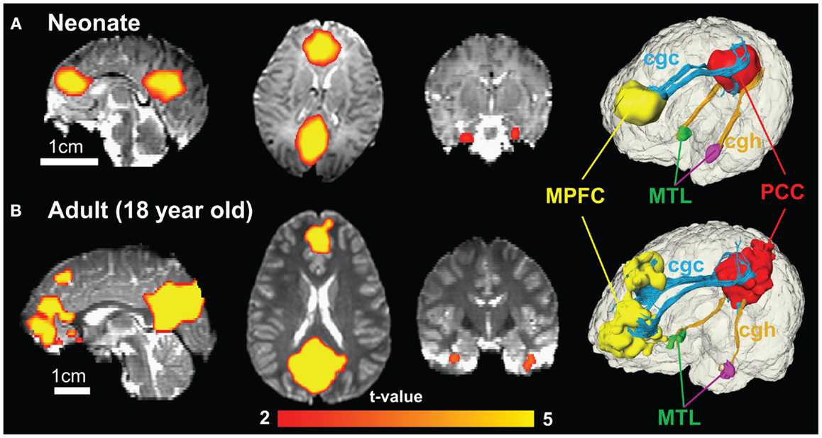
So do you make up for lost cognitive flexibility time for children? Or for yourself as a person of structure and routine? The answer is no. It is more important to make active choices to be exposed to the internal and external worlds that are immediate and to ensure that physical movements are consciously added in a 24 hour period than to make up for the over a year of standing still. Scientific studies have isolated the executive functions that aim at cognitive flexibility, which include the abilities to shift one’s thinking (flexibility), updating the learning that has been made based on the thinking shift (working memory), and response inhibition. In Uddin’s 2021 study, Cognitive and behavioural flexibility: neural mechanisms and clinical considerations she explains the core processes in thinking flexibility with this figure:

Fig. 1

Fig. 1: Core cognitive processes and brain network interactions underlying flexibility in the human brain. From: Cognitive and behavioural flexibility: neural mechanisms and clinical considerations

These brain maps were established with the use of automated meta-analyses of published functional neuroimaging studies can be conducted with Neurosynth, a Web-based platform that uses text mining to extract activation coordinates from studies reporting on a specific psychological term of interest and machine learning to estimate the likelihood that activation maps are associated with specific psychological terms, thus creating a mapping between neural and cognitive states. In the study, Neurosynth reveals that brain imaging studies including the terms ‘shifting’, ‘updating’ and ‘inhibition’ report highly overlapping patterns of activation in lateral frontoparietal and mid-cingulo-insular brain regions, underscoring the difficulty of isolating the construct of flexibility from associated executive functions.

This means that cognitive flexibility is an activity that requires the whole brain, and if that is the case, then it requires a complete human experience. In an article by Sahakian, et. al in the World Economic Forum site called, Why is cognitive flexibility important and how can you improve it? they indicate that Cognitive flexibility provides us with the ability to see that what we are doing is not leading to success and to make the appropriate changes to achieve it. Flexible thinking is key to creativity – in other words, the ability to think of new ideas, make novel connections between ideas, and make new inventions. It also supports academic and work skills such as problem-solving.

They also write that cognitive flexibility can also help protect against a number of biases, such as confirmation bias. That’s because people who are cognitively flexible are better at recognizing potential faults and difficulties in themselves and using strategies to overcome these faults. See their table below showing the flexibility representations:

How do we become adept at choosing to be flexible especially in situations that give little determination of what we can control? Aside from practicing the principles of evidence-based psychological therapy which allows people to change their patterns of thoughts and behavior (Cognitive Behavioral Therapy or CBT), Structure learning has been proven to be potentially another way. It has been described as a person’s ability to extract information about the structure of a complex environment and then decipher initially incomprehensible streams of sensory information via the process of elimination. This specific type of learning taps into the similar frontal and striatal brain regions as cognitive flexibility, thus exposure and practice are the keys to successful learning.

Go forth, be human, and explore!

Advertisement

Response Inhibition in the 21st Century (EF Skills Series)

If there is a human condition that is written most about second to human emotions, it would be that of impulse. Motivation powered by Dopamine and sped by Acetylcholine (AcH) is not necessarily supported by the higher-order skills that the Frontal Cortex is known for when impulse gets in the way. For example, when a choice is made by a person to eat what is body assistive or eat what is fast and palatable, chances are impulses drive the attention toward the sensory appealing choice.

It is by responding with delayed gratification, to not take the most convenient method to an end goal, that we can assess the process of each step toward the goal. How then can pacing be valued equitably when it is the efficiency of speed and productivity that pays higher dividends? What was pioneered by the Marshmallow test conducted by Walter Mischel in 1972 on preschoolers continues to be replicated, and bottom-line conclusions are the better a person redirects and controls his or her impulses, the clearer the direction of the path toward the end goal/s. Though not entirely specified, the original test was an exercise of concretizing “waiting” into something that had a “reward” in the end, and that the Marshmallow itself was not the real goal.

The ultimate reward of response inhibition is to process the game of waiting. Waiting on public transportation with a throng of people. Waiting and completing the morning routine despite sleep deprivation. Sticking to the regimen that is pro-health (physical, mental, emotional). To play the game of waiting efficiently, strategies that are at the person’s disposal are in the Inhibitory control toolkit, including scenario playing such as Stimulus–Stimulus- and Stimulus–Response-conflict with the many selves and environments a person interacts with.

The Waiting Game can be very difficult for those who have challenges attending to simple steps. The hippocampus is unable to maintain bridges with the Pre-Frontal Cortex (PFC) when the thoughts are not in sync, where working memory is affected since inhibiting responses allow memories to be processed clearly. Mindfulness exercises do increase the strengthening of neural pathways from the parietal cortex to the frontal cortex as it supports the just ‘being’ state without conclusions. However, as with attention, mindfulness requires a push-pull of one’s attentional inhibition, which means the decision to let some things just pass by like clouds while maintaining focus on the journey of the mind during the session is multilayered. Do I respond to the thought cloud of the ding of the phone, maybe that was an email…or do I cut this short, dinner time is close and I have friends coming later…wait, what was I supposed to be concentrating on…?

The yin-yang of inhibitory control behavior has many names in the field of behavioral science. Response Inhibition is also referred to as ‘Behavioral Inhibition,’ ‘Motor Inhibition,’ ‘Prepotent Response Inhibition,’ and ‘(Attention) Restraint’; while Attentional Inhibition is also referred to as ‘Interference Control,’ ‘Interference Suppression,’ ‘Resistance to (Distracter) Interference,’ and ‘Attention Constraint.’ Think of the two as the demonstration of Newton’s third law is: For every action, there is an equal and opposite reaction. This means response and attention in every interaction are the pair of forces acting on the two interacting choice objects.

Both these inhibition rubrics are necessary when interacting with general life situations that require working memory (both in verbal and auditory means), visual memory (stop and analyze then respond), reflexive responses (go/no-go tasks and responses), word spatial recognition, and figure grounding contrast and comparative analysis. In short, in every single aspect of living that requires a decision between options, it is the brain’s processes that attach to memories and emotions that decide between the choice to stay the course and inhibit or choose the one that interferes with the course and give in to the attentional distracter.

According to Verbruggen, et al (2014) in their study, The Inhibitory Control Reflex, performance in response inhibition paradigms can be modeled as an independent “horse race” between a go process, which is triggered by the presentation of a go stimulus, and a stop process, which is triggered by the presentation of the no-go stimulus or the stop signal. When the stop process finishes before the go process, response inhibition is successful and no response is emitted (signal-inhibit); when the go process finishes before the stop process, response inhibition is unsuccessful and the response is incorrectly emitted (signal-respond). The latency of the stop process (stop-signal reaction time or SSRT) is covert, but it can be estimated in the stop-signal task. SSRT has proven to be an important measure of the cognitive control processes that are involved in stopping.

Yet it is the weight of the end goal that determines the type of inhibitory control that one ultimately uses more of. And as mentioned earlier, the more concrete the process of inhibition by the fruition of an actual marked goal, the more likely one stays the course and avoids attentional constraints. In order to facilitate goal-directed behavior, the proper motor actions in a given context need to be selected and then executed. Regions within the medial wall of the
frontal cortex, in particular, the supplementary motor area (SMA), have been implicated in response preparation, selection, and execution. Single-cell recordings in monkeys performing a delayed reaction task reveal a distinction between the rostral portion of the SMA (‘‘pre-SMA’’) and the caudal portion of the SMA (‘‘SMA proper’’); neuronal activity is seen in the SMA proper only during the response; activity in the pre-SMA, however, is associated with both the delay period and the response, suggesting that the pre-SMA is involved in both preparation and selection of the response. This means that inhibitory responses are best strengthened when paired with a visual spatial or perceptual motor component prior to perceiving and selecting what the response selection is.

This could also support the idea that the younger the developmental age, the more multi-modal means are necessary to train the inhibitory models of the PFC. Competing with the gadgets that provide instant gratification however is much more prevalent since coming out of the lockdown, making the responses less precise and more impatient. Also part of the multi-modal means is to create a fruitful digital diet, one that is realistic and supportive of the ‘school or home work’ that still needs to be done in the platform while being able to segregate the ‘fun work’ that is embedded in the devices. One possibility is that increased functional segregation of neural networks during development results in greater differentiation of individual inhibitory control mechanisms at the behavioral level. Evidence for this is suggested by findings from the study done by Mostofsky, et. al (May 2021) Response Inhibition and Response Selection: Two Sides of the Same Coin) have found that tasks related to executive control tend to coalesce as a single factor in young children, but form a multi-factor construct in adults. This does not mean however that the addictive properties of smart devices do not affect adults the way they do children; response inhibition is key for moderation and for caudate nucleus sustainability, one of the social centers of the brain.

A Review on Brain’s Health

As much as there have been many suggestions and studies on how to cope with the pandemic using multi-modal means, such as apps, vitamins or physical exercise, or even new hobbies and food adventures. And that is on the upside of things. Coping skills when the cards are stacked against your favor hyper focuses on survival from moment to moment when the brain is overloaded and tasked with overchoice.

Multi-million-dollar industries have been built on people’s pursuits on the fountain of youth and keeping the brain young or at least to match the youthful vigor of how the body should feel. Emotionally despite our wanting for vigor, we are brought back to realization by our brain’s level of resources or how to retrieve it as the amygdala automatically pushes past the suggestion of pain or difficulty when basic needs are of the essence.

Caring for the brain’s emotions are interdependent on the human capacity for social cognition when reading body language and expressions. And in the stressful pandemic times, adaptive social behavior and mental well-being also require inferring the absence of emotional content. Of relevance during exceptional events such as the coronavirus disease 2019 (COVID-19) pandemic involving social distancing and isolation as well as higher levels of anxiety but also, requiring intact social communication and empathy. (Sokolove et al, 2020)

When one is required to maintain social distance and/or uses PPE while interacting with another, the interplay between the amygdala and insula is elevated as it contributes substantially to the processing of social signals that convey emotionally neutral information, underwriting a fundamental yet underestimated component of adaptive social cognition and behavior. The absence of stimulatory emotion presents a difficulty of processing in the bond communication between the amygdala and insula with the ability to infer the
absence of emotion in body language, including with the processing within the amygdala and insula have been associated with emotion perception. Ultimately, when caring for the brain’s health scanning other people’s words and delivery are what is needed for homeostasis.

So, the brain begins searching for alternates in reading and writing ’emotion’ in times of crises to maintain itself. Depending on the coping mechanisms embedded within the connections, physical and/or psychological experience that binds with an external event, this one being the lack of neurotypical emotion perception could potentially ripple into pre-existing trauma. There are growing indications that these trauma-exposed individuals show
neural alterations, even in the absence of clinical symptoms and tasks requiring the regulation of emotions and impulses seem to be particularly affected. Sabrina Golde et.al

studied 25 women (mean age 31.5 ± 9.7 years) who had suffered severe early life trauma but who did not have a history of or current psychiatric disorder, with 25 age- and education-matched trauma-naïve women by utilizing fMRI study investigated response inhibition of emotional faces before and after psychosocial stress situations in 2019. In both groups, nontraumatic stress significantly impaired response inhibition of fearful facial expressions but did not affect inhibition of happy or neutral stimuli. When inhibiting responses to fearful faces under stress, women who had experienced severe and multiple traumatic events before the age of 18 demonstrated a decreased brain signal in the left inferior frontal gyrus in the frontal lobe, but a marginally increased signal in the right anterior insula (between the frontal and temporal lobes) compared to trauma-naïve control subjects.

They also discovered that in trauma-naïve control subjects only, the inferior frontal gyrus activation was linked to a lower rate of stress-induced false alarms, whereas in women who had experienced severe and multiple traumatic events before the age of 18, insula activation was linked to a higher number of false alarms.

Of course, that begs the question, would brain health be different between a male brain in comparison to the female brain? Are brains not simply just as generic as other parts of the body such as the skeletal and muscular systems? Would the same study above if applied to men render equitable results as the women have displayed? Perhaps, as brain function is dependent on synapses and stimuli response. However as unique as every individual is, the responses can and will be varied according to the emotional pairing, regardless of assigned biological sex.

After all neuroplasticity happens to people, mostly in the background, positive or negative. The learning experience is constantly evolving, including in what is automatic such as driving, to the more complex tasks such as learning a new skill or trade. Yet in the viewpoint from a healthy brain, neuroplasticity would support neural pruning at every stage of life: keeping only the connections that are meaningful and consistently used for an end goal while allowing brain real estate to grow for additional new experiences to bind. If, however the exposure is constantly negative neuroplasticity such as death, destruction, or violence, the brain’s stress levels are increased thus decreasing overall expanded connections and brain weight.

Sandra Racionero-Plaza, et al., found in their research article, “Architects of Their Own Brain.” Social Impact of an Intervention Study for the Prevention of Gender-Based Violence in Adolescence sought to review the social impact of psychology in the field of teen gender violence from participants in the MEMO4LOVE project comprised 126 adolescents attending three different high schools in Barcelona (Spain). The aim was to report on the potential social impact achieved by an intervention study consisting of seven interventions on the preventive socialization of gender violence, with specific interventions focused on seven different topics. Part of the intervention were dimensions of the influence of the dominant coercive discourse in adolescents’ sexual-affective life, accompanied by egalitarian dialogue with the adolescents on how to break free from such discourse and thus avoid violence in sporadic or stable sexual-affective relationships. Below is what they found:

1) The adolescents who took part in these interventions reported developing critical consciousness regarding the presence of the dominant coercive discourse in society and in their lives. The greater the transformation of peer interactions, the greater the prevention of gender-based violence victimization.

2) It is necessary that intervention strategies in education provide adolescents with tools that enable them to be more critical about violence in society. Interventions provided them with cognitive tools to better analyze their own and others’ sexual-affective thinking, emotions, and behaviors in ways that favor the rejection of violent males and violent sexual-affective relationships

3) Change in preferences and attraction, with greater rejection of violent men and enhanced attraction toward egalitarian masculinities. It is significant to be aware of the dominant coercive discourse, but what is even more fundamental is to change the response of attraction toward men with violent attitudes and behaviors; that is what will make a difference in terms of preventing gender-based violence victimization.

Society therefore continues to impact the neuroplasticity quality with our degrees of interaction with it. Aside from gender and age groups, it is of significance to mention that brain health across races also have complex layers affecting neuroplasticity. In the research article, Insights From African American Older Adults on Brain Health Research Engagement: “Need to See the Need” Soshana Bardach, et al., posited that African Americans (AAs) have an elevated risk of developing dementia, yet are underrepresented in clinical research. In order for the 21 AAs aged 35-86 years to agree to be part of the study, the researchers had to ensure that the facilitators were AA from their community, laying out clearly and plainly the methodology that would be used to gather data which is called Photovoice, and to be transparent about the reasons behind the data gathering.

The use of Photovoice in data collection is a community-based, participatory-action research methodology, centered on the premise that sharing perceptions and experiences through photography and discussion leads to an awareness and understanding of relevant issues (Harley et al, 2015) It was through this method that the discussion of brain health research among AAs highlighted several barriers to participation and corresponding strategies for addressing the identified barriers and encouraging engagement. The barriers and solutions were in the areas of (a) mistrust stemming from historical mistreatment and reinforced by contemporary injustices, (b) avoidance and fear of acknowledging problems such as being afraid of what they may find out or not wanting to know as barriers to seeking a diagnosis, and (c) seeing the risks of research, but not the need as they anticipate potential unpleasant aspects of research participation were more tangible than the benefits.

This then brings us to consider that brain health is not only a result of positive neuroplasticity but a contiguous history of sustainable emotions stemming from society and communities. The deterioration of the brain affected by genetics is also sped up by neurochemicals that inflame the stress response on a consistent basis thus making recovery to a biologically sound baseline unattainable. Anti-declining the brain, simply put, is to bring it to a social balance. An immediate way is to encourage pro-social behaviors between humans and/or animal species. Pro-social behaviors—actions that help others, often without regard to whether they directly benefit us—are vital for social cohesion. (Lockwood, et al, 2020) The idea is that to do acts that benefit other people, we first must learn what actions in the world will lead to something rewarding and positive happening to them, all processed in the anterior cingulate cortex of the brain. When we perform an action, and it results in a reward for another person, it is easy to imagine ourselves in their shoes and vicariously experience what it would be like to get a similar reward ourselves.

If there are limited opportunities for vicarious learning or pro-social behaviors, such as when in a global crises, how can balance be achieved? Ironically, Polyphenol-Rich Interventions are in order to influence the sensitivity of natural neurotransmitters and adjust levels naturally. Polyphenol compounds, such as flavonoids, phenolic acids and tannins, are found in varying concentrations in a range of plant-based food sources (e.g., legumes, fruit, vegetables, herbal extracts, spices, coffee, tea and cocoa), and their effects on human health have drawn considerable attention. (Ammar, et al 2020). It is the self-socializing method by virtue of self-care that supports choices of the quality of brain health. At this particular juncture, the use of the human amygdala is its response to other emotionally significant stimuli beyond threat, including positive or reward information, but also novelty and non-emotional salient stimuli with personal impact or goal-related significance. This diversity of response patterns has led to recent theoretical accounts proposing that the amygdala may actually encode the “relevance” of events, which is determined by the goals, needs, or values of the individual, in a context-dependent manner. (Guex et al, 2020) This account accords with psychological theories, such as the component process model of emotional appraisal as would in patterns of self-socialized self-care.

The anterior cingulate cortex and the amygdala would need support when communicating emotions-bound signals to each other. It is with the subcortical structures of the basal ganglia (BG) and the cerebellum that support multiple domains including affective processes such as emotion recognition, subjective feeling elicitation and reward valuation. By regulating
cortical oscillations to guide learning and strengthening rewarded behaviors or thought patterns to achieve a desired goal state, these regions can shape the way an individual processes emotional stimuli. (Pierce J, et al 2020) In essence, no matter how much objective data could be presented to standardize the ideal brain health for a given time frame, developmental stage, race or gender, the conclusion will rest upon every individual’s experience or norm of how their brains are in status quo or if they are on polar opposites. Whatever the case may be, brain health is essential for quality of life.

Electromagnets and the Servings of Hope

So got the latest iPhone and accessories? That will definitely speed productivity and social connections. Do you have children who are electronically savvy with these devices? Depending on who is doing the research, there may be a mixed bag of OOOHS and OH NOOOs.

EMF1Here we explain. Most of our speedy, high tech devices are powered by Electromagnetic Fields (EMFs). Cindy Sage, MA, and Nancy Evans, BS explain in their handout prepared for a website called Healthy Schools in 2011 in detail the kinds of EMFs that we encounter everyday:

Extremely low frequency electromagnetic fields (ELF-EMF) are generated from appliances and other items that  use electricity (power frequency fields).

Radiofrequency (RF-EMF) is generated by wireless technologies such as cellular and cordless phones.

“Dirty electricity” is a term used to describe low kilohertz frequency fields that can be thought of as an unintentional RF pollutant on electrical wiring and into living space. Power is “dirty” or polluted when it contains the high frequency signals flowing through overloaded wires, and not just the clean 60 Hz power that’s created at the source.

We are all aware of the benefits of modernization and upgrading to the latest gadgetry. We are able to cram as much work/leisure/information as possible in the shortest amount of time. It improves productivity, increases quantity of life skills, and promotes connectivity only science fiction writers used to dream about.  Ironically (good or bad), in 2010 MIT neuroscientists have now shown they can influence those judgments by interfering with activity in a specific brain region — a finding that helps reveal how the brain constructs morality. The researchers, led by Rebecca Saxe, MIT assistant professor
of brain and cognitive sciences disrupted activity brain region known as the right temporo-parietal junction (TPJ) by inducing a current in the brain using a magnetic field applied to the scalp.  The researchers used a noninvasive technique known as transcranial magnetic stimulation (TMS) to selectively interfere with brain activity in the right TPJ. The magnetic field applied to a small area of the skull creates weak electric currents that impede nearby brain cells’ ability to fire normally, but the effect is only temporary.

They found that the subjects’ ability to make moral judgments that require an understanding of other people’s intentions was impaired. The researchers believe that TMS interfered with subjects’ ability to interpret others’ intentions, forcing them to rely more on outcome information to make their judgments.

So EMFs literally can assist in changing our minds, literally. How about our health? And our young people’s development?

EMF2
A report commissioned by T-Mobile and Deutsche Telecom MobilNet GmbH prepared in 2000 reviews effects such as gene toxicity, cellular processes, effects on the immune system, central nervous system, hormone systems and connections with cancer and infertility. This was utilized by the Commonwealth Club of California’s Program on Health Effects of Cell Phones, Wireless Technologies & Electromagnetic Fields With Leading Experts in November 2010.

In their study, Dr Kerstin Hennies, Dr H.‐Peter Neitzke and Dr Hartmut Voigt in behalf of the Telecom companies found:

1. Given the results of the present epidemiological studies, it can be concluded that electromagnetic fields with frequencies in the mobile telecommunications range do play a role in the development of cancer. This is particularly notable for tumours of the central nervous system, for which there is only the one epidemiological study so far, examining the actual use of mobile phones.

2. Damaging effects on the immune system which can aid the development of illnesses as demonstrated higher secretions of stress hormones in humans.

3. Effects of high frequency electromagnetic fields on the central nervous system are proven for intensities well below the current guidelines.

4. The terms ‘electrosensitivity’ or ‘electromagnetic hypersensitivity’ describe disturbances of well‐being and impairments of health, such as they are suffered by certain sensitive people when working with or being in the presence of devices and equipment emitting electrical, magnetic or electromagnetic fields.

They also conclude: “A particular problem in this exposure group is posed by children and adolescents, not only because their organism is still developing and therefore particularly susceptible, but also because many cp-radiationadolescents have come to be the most regular users of mobile phones. Advertising towards this population group should be banned. Furthermore, particular efforts should be made to lower the exposures during calls. It would be recommendable to conduct (covert) advertising campaigns propagating the use of headsets. It would also be important to develop communications and advertising aiming at minimising the exposures created by carrying mobile phones in standby mode on the body.”

That was in 2000. That is not the case in 2015. Covert would not be the word for the in-your-hand ads aimed to the youngest demographic possible (e.g. no more teen data overages…hint hint). So what to do?

Here’s the practical, scientific approach recommended by experts: Use a corded phone (land line) as your regular telephone. If you need to use a cordless phone or cell phone, use a headset (wired only) whenever possible and/or use your phone on speakerphone. Text rather than talk. Keep your calls very brief, and hold your cell phone away from your head and body, especially when the phone is connecting your call. Children should not use cell phones or cordless phones. Studies show children have a five-fold risk of malignant brain tumors in a shorter time than adults. 

hope1The other recommendation? Healthy servings on Hope. The brain on hope supports a growing body of scientific evidence that points to the conclusion that optimism may be hardwired by evolution into the human brain. The science of optimism, once scorned as an intellectually suspect province of pep rallies and smiley faces, is opening a new window on the workings of human consciousness. What it shows could fuel a revolution in psychology, as the field comes to grips with accumulating evidence that our brains are constantly being shaped by the future.

Findings from a study  conducted a few years ago with prominent neuroscientist Elizabeth Phelps and Tali Sharot suggest that directing our thoughts of the future toward the positive is a result of our frontal cortex’s communicating with subcortical regions deep in our brain. The frontal cortex, a large area behind the forehead, is the most recently evolved part of the brain. It is larger in humans than in other primates and is critical for many complex human functions such as language and goal setting.

Using a functional magnetic resonance imaging (fMRI) scanner, the researchers recorded brain activity in volunteers as they imagined specific events that might occur to them in the future. Some of the events were desirable (a great date or winning a large sum of money), and some were undesirable (losing a wallet, ending a romantic relationship). The volunteers reported that their images of sought-after events were richer and more vivid than those of unwanted events.

This matched the enhanced activity observed in two critical regions of the brain: the amygdala, a small structure deep in the brain that is central to the processing of emotion, and the rostral anterior cingulate cortex (rACC), an area of the frontal cortex that modulates emotion and motivation. The rACC acts like a traffic conductor, enhancing the flow of positive emotions and associations. The more optimistic a person was, the higher the activity in these regions was while imagining positive future events (relative to negative ones) and the stronger the connectivity between the two structures.

The positive physiological effects of hope are well-documented, most recently by CNN in 2013  in Jerome Groopman’s “The Anatomy of Hope,” where he writes: “Researchers are learning that a change in mind-set has the power to alter neurochemistry.”  His research also showed that during the course of illness, belief and expectation have an impact on the nervous system which, in turn, sets off a chain reaction that makes improvement and recovery more likely. Groopman observed that hope does not just involve a mind-to-body connection, but also a body-to-mind connection, where neural input about one’s physical condition serves as a moderator of positive and negative emotions.

hope2Shane Lopez, author of the new book “Making Hope Happen,” believes hope is the stuff of change, recovery and healing. Hope is half optimism, Lopez explains. The other half is the belief in the power that you can make it so.There is a profound difference between hoping and wishing, he continues. Wishing encourages passivity, whereas hope represents an active stance.

“Wishing is the fantasy that everything is going to turn out OK. Hoping is actually showing up for the hard work.”

And it is hard work to find moderation between technological use and traditional, generalist methods of living. A line needs to be drawn for generations after us to have a chance at a future before they can manipulate it, or else all the forward thinking and efficiency cramming we did in our heyday for them is mismatched and misaligned. Balancing between picking up a book with pages AND including one or two websites for research creates a nifty scale bridging the survival rate of the future and wisdom from longevity of the past.

Silence.

Silence2Think of one place in the world that is still completely silent. Deep ocean perhaps? That must still be relatively silent. Is there any country in the world that is silent?

In 2011, the Finnish Tourist Board released a series of photographs of lone figures in the wilderness, with the caption “Silence, Please.” An international “country branding” consultant, Simon Anholt, proposed the playful tagline “No talking, but action.” And a Finnish watch company, Rönkkö, launched its own new slogan: “Handmade in Finnish silence.”

“We decided, instead of saying that it’s really empty and really quiet and nobody is talking about anything here, let’s embrace it and make it a good thing,” explains Eva Kiviranta, who manages social media for VisitFinland.com.

So Finland 4 years ago decided to capitalize on their silent status seeing that the world thrives on noise. However on the scientific front, the sound of silence stimulates a bigger response in the brain of a listener than music itself.  According to research, silence provides intriguing support for modern composers who put as much emphasis on a lack of sound as sound itself.

Led by Dr. Vinod Menon of Stanford University School of Medicine, the researchers published their findings in 2010 in the journal Neuron. The team showed that music engages the areas of the brain involved with paying attention, making predictions and updating the event in memory.

In what has to be one of the most pleasant brain studies on record, researchers asked subjects to listen to symphonies by the 18th century English baroque composer William Boyce, chosen because they are relatively short and comprise well-defined movements, punctuated by pauses.

As the subjects listened the researchers scanned their brains using functional magnetic resonance imaging. This widely used technique involves using harmless magnetic fields and radio waves to measure blood flow in brain regions, which reveals the amount of activity in those regions.

But while music may soothe the savage beast, the brain responds efficiently to the sound of silence. Peak brain activity occurred during a short period between musical movements, when seemingly NOTHING was happening. Two distinct networks of brain regions on the right side of the brain were involved, suggesting it was excited at the anticipation of more to come, or predicting the next movement.

“Our study suggests that silence, appropriately structured, is a device that allows the composer to achieve certain goals,” Dr. Menon told The Daily Telegraph. “It can arrest the listener’s attention and create conditions that facilitate anticipatory processes related to previously heard sequences of sounds. I suspect that silences inserted by composers such as Stockhausen and Philip Glass can elicit intense physiological and brain responses, although how long these responses can be sustained and manipulated is not yet clear. Modern brain imaging techniques might help to clarify how silence can be manipulated to create novel musical experiences.” Silence1

Karlheinz Stockhausen, a German musical composer discussed these experiences, for example in one composition where there are silences up to about one minute. “I discovered a new way to prepare for a certain duration of silence by what happens just before the silence, so that one can hear again, like an echo, the figures or structures before the silences. I think there is a very secret science of musical composition in knowing what one has to do before a silence in order to make the following silence meaningful.”

A team of University of Oregon researchers isolated an independent processing channel of synapses inside the brain’s auditory cortex that deals specifically with shutting off sound processing at appropriate times. Such regulation is vital for hearing and for understanding speech.

The discovery, found in the Feb. 2010 issue of the journal Neuron, goes against a long-held assumption that the signaling of a sound’s appearance and its subsequent disappearance are both handled by the same pathway. The new finding, which supports an emerging theory that a separate set of synapses is responsible, could lead to new, distinctly targeted therapies such as improved hearing devices, said Michael Wehr, a professor of psychology and member of the UO Institute of Neuroscience.

To do the research, Mr. Wehr and two UO undergraduate students — lead author Ben Scholl, then a graduate student at the Oregon Health and Science University in Portland, and Xiang Gao — monitored the activity of neurons and their connecting synapses as rats were exposed to millisecond bursts of tones, looking at the responses to both the start and end of a sound. They tested varying lengths and frequencies of sounds in a series of experiments.

“It looks like there is a whole separate channel that goes all the way from the ear up to the brain that is specialized to process sound offsets,” Mr. Wehr said. The two channels finally come together in a brain region called the auditory cortex, situated in the temporal lobe.

The research team also noted that responses to the end of a sound involved different frequency tuning, duration and amplitude than those involved in processing the start of a sound, findings that agree with a trend cited in at least three other studies in the last decade.

It became clear with the findings that one set of synapses responded “very strongly at the onset of sounds,” but a different set of synapses responded to the sudden disappearance of sounds. There was no overlap of the two responding sets, the researchers noted. The end of one sound did not affect the response to a new sound, thus reinforcing the idea of separate processing channels.

“Being able to perceive when sound stops is very important for speech processing,” Mr. Wehr said. “One of the really hard problems in speech is finding the boundaries between the different parts of words. It is really not well understood how the brain does that.”

As an example, he noted the difficulty some people have when they are at a noisy cocktail party and are trying to follow one conversation amid competing background noises. “We think that we’ve discovered brain mechanisms that are important in finding the necessary boundaries between words that help to allow for successful speech recognition and hearing,” he said.

Mandy-Hale-Silence-QuotesThe research conducted aimed to provide a general understanding of how areas of the brain function. These findings could also prove useful in working with children who have deficits in speech and learning, as well as in the design of hearing aids and cochlear implants. He also noted that people with dyslexia have problems defining the boundaries of sounds in speech, and tapping these processing areas in therapy could boost reading skills.

In the course of developing the human capacity, movement and noise require a cease and desist button for either of them to matter in the first place. In spite of the brain’s 24/7 processes, most of them we actually only experience, not hear. Thus the point of silence — it is the definition of a fulfilled experience that personalizes what otherwise would be lost in the noise.

Learning’s Many Costs, First Quarter 2015 Edition

Flipping channels, Dr. Phil comes onto the main screen and has a mother and daughter in opposite angles to him. The problems seemed to be typical psychological, reality-show formula: mom is overprotective of her teenage daughter, teenage daughter ‘rebels’ and begins a long distance relationship with a man 10 years her senior (who also happens to be a drug dealer still serving time), and the conflicts between them become unbearable. Dr. Phil in his infinite wisdom brought out his objectivity to the mother, and then to the daughter. It is however the explanation to the daughter that resonates with her: “Now remember, it is the Neocortex that develops last in the brain, and that means right now, you’re not able yet to have complete insight to all of the consequences of your actions. This is why you would need guidance with some of your decisions, including starting a relationship with a currently jailed drug dealer.”

Most of us have a familiarity with family drama played out on television.  Whether the purpose is to entertain or to educate, the family’s dysfunctional dynamic from one to the next seem to be rooted deep into social, educational and economic factors. How then is the brain development affected? Is Dr. Phil’s approach by combining the brain science with family dynamics warranted?

money-house

In the early online edition of the journal Nature Neuroscience (March 30, 2015), investigators from nine universities across the country reports correlative links between family income and brain structure. Relationships between the brain and family income were strongest in the lowest end of the economic range, thus suggesting that interventional policies aimed at these children may have the largest societal impact. The study, led by researchers at The Saban Research Institute of Children’s Hospital Los Angeles and Columbia University Medical Center.

In the largest study of its kind to date, the researchers looked at 1,099 typically developing individuals between the ages of 3 and 20 years as part of the multi-site Pediatric Imaging, Neurocognition and Genetics (PING) study. Associations between socioeconomic factors (including parent education and family income) and measurements of surface area of the brain were drawn from demographic and developmental history questionnaires, as well as high-resolution brain MRIs. Statistics (controlled for education, age and genetic ancestry)  showed that income was nonlinearly associated with brain surface area, and that income was more strongly associated with the brain than was parental educational attainment.

First author Kimberly G. Noble, MD, PhD says, “Specifically, among children from the lowest-income families, small differences in income were associated with relatively large differences in surface area in a number of regions of the brain associated with skills important for academic success. ” Dr. Noble is an assistant professor of pediatrics and director of the Neurocognition, Early Experience and Development (NEED) Lab of Columbia University Medical Center. She is also an associate professor of Neuroscience and Education at Teachers College, Columbia University.

Conversely, among children from higher-income families, incremental increases in income level were associated with much smaller differences in surface area. Higher income was also associated with better performance in certain cognitive skills; cognitive differences that could be accounted for by greater brain surface area.

“While in no way implying that a child’s socioeconomic circumstances lead to immutable changes in brain development or cognition, our data suggest that wider access to resources likely afforded by the more affluent may lead to differences in a child’s brain structure,” said Elizabeth Sowell, PhD, director of the Developmental Cognitive Neuroimaging Laboratory, part of the Institute for the Developing Mind at CHLA.  “Family income is linked to factors such as nutrition, health care, schools, play areas and, sometimes, air quality,” added Dr. Sowell, indicating that everything going on in the environment shapes the developing brain. “Future research may address the question of whether changing a child’s environment — for instance, through social policies aimed at reducing family poverty — could change the trajectory of brain development and cognition for the better.”

From the socio-economic factors, we take a look at socio-psychological factors, some of which affect our ability to create meaningful connections. A recent study from the University of Georgia shows differences in brain structure according to how trusting people are of others.

The team of researchers used two measures to determine the trust levels of 82 study participants. The participants filled out TRUSTING CHILDa self-reported questionnaire about their tendency to trust others. They also were shown pictures of faces with neutral facial expressions, and were asked to evaluate how trustworthy they found each person in the picture. This gave researchers a metric, on a spectrum, of how trusting each participant was of others.

Researchers then took MRI scans of the participants’ brains to determine how brain structure is associated with the tendency to be more trusting of others. What researchers found were differences in two areas of the brain.

“The most important finding was that the grey matter volume was greater in the ventral medial prefrontal cortex, which is the brain region that serves to evaluate social rewards, in people that tended to be more trusting of others,” said the study’s lead author Brian Haas, an assistant professor in the department of psychology.

“Another finding that we observed was for a brain region called the amygdala. The volume of this area of the brain, which codes for emotional saliency, was greater in those that were both most trusting and least trusting of others. If something is emotionally important to us, the amygdala helps us code and remember it.”

The long term hope for the research may have implications for future treatments of psychological conditions such as autism. Future studies may focus on how, and if, trust can be improved and whether the brain is malleable according to the type of communication someone has with another. “There are conditions, like autism, that are characterized by deficits in being able to process the world socially, one of which is the ability to trust people,” Dr. Haas said. “Here we have converging evidence that these brain regions are important for trust; and if we can understand how these differences relate to specific social processes, then we may be able to develop more targeted treatment techniques for people who have deficits in social cognition.”

So what can we do as a community with fragile families who have young children in such a fast-paced, competitive, and digitally plugged world? Begin the developmental awareness young for a firmer foundation with research-backed programs.

Supporting this is new research from UNC’s Frank Porter Graham Child Development Institute (FPG) revealing high-quality early education is especially advantageous for children when they start younger and continue longer. Not only does more high-quality early education significantly boost the language skills of children from low-income families, children whose first language is not English benefit even more.

“These findings show that more high-quality early education and care can narrow the achievement gap before children reach kindergarten,” said Noreen M. Yazejian, principal investigator of FPG’s Educare Learning Network Implementation Study. “Children from low-income families can improve their standing relative to their middle class peers.”

Ms. Yazejian said previous research has shown language skills are most malleable for children before age 4, which in large part explains high-quality early education’s powerful effects. Her study examined children’s receptive language skills–the ability to hear and understand words–because these particular skills are an excellent predictor of later academic success.

According to Yazejian, Educare classrooms offered the chance to study children enrolled in high-quality early education and care from the earliest ages. Educare is an enhanced Early Head Start and Head Start program for low-income, high-needs children from 6 weeks old until entry into kindergarten. The model has been replicated in 20 schools nationwide over the last 15 years.

“Educare’s comprehensive approach to early childhood education aims to level the playing field for children living in poverty,” said Portia Kennel, executive director of the Educare Learning Network. “This new study confirms that we need to include the earliest years of life as part of our nation’s education system. Quality early education prepares vulnerable children for success by preventing the achievement gap that appears long before kindergarten.”

Many people traditionally have viewed early care for infants primarily as a support for mothers who want to work and not as an essential component of early schooling. However, findings from the FPG study add to a growing body of research revealing better outcomes for children from low-income families who start high-quality education earlier and stay in it longer.

Earlier research has shown the English language skills that dual-language learners develop prior to kindergarten can predict educational achievement through eighth grade, but keeping skills in the home language also is beneficial. Home language skills are related to long-term social, emotional, cognitive, and academic outcomes.

“Most dual-language learners in this study were in classrooms where English was the primary instructional language but in which one staff member could use their home language as needed to support learning,” Ms. Yazejian explained. “It’s not surprising our findings show they quickly acquired skills in English. That’s why it’s reassuring that our study found that the acquisition of English language skills in Educare classrooms does not come at the expense of Spanish skills.”

The number of young children who speak a language other than English at home is growing, and this study contributes valuable new information to the field. “It’s encouraging to see that dual-language learners are making strides that form the critical foundation for later learning,” according to Ms. Kennel.

Ms.Yazejian encourages the thinking that more than one year of high quality early care and education brings greater benefits for children. “The differences we found in this study, extrapolated to thousands of children–especially dual-language learners–could add up to lasting effects and lower public education costs.”

cropped_77388_Royalty_Free_RF_Clipart_Illustration_Of_A_Diverse_Group_Of_Children_Spelling_The_Word_Preschool1

T’is the Season for Depression, And How to Shake it Off

It is definitely the heart of winter, mid-February, and we would be lucky if we had a week that didn’t have frigid temperatures, snowfall and blizzard-type winds, and long nights. Compared to the spring and summer seasons, winter in the northeastern hemisphere of the United States challenge even the happiest of people — weather conditions force us to stay indoors as much as possible, unless we emancipate ourselves from the city during these times into a cabin in the woods with hot cocoa and recreational activities meant for the season.

I am willing to bet however we aren’t all as lucky; city life and the responsibilities that come with it keep us close to it even in the harshest winter weather conditions. Compound that feeling of being trapped with heavy layered clothing, dangerous walking and commuting conditions, and gray, dreary short days that at 5 pm remind you it is wintertime with it’s pitch black quick nightfall.

Understandably this time of year, after the holidays have passed, depression sets in full force. There are those of us, however, who suffer a more consistent, cyclical type of depression this time of year called Season Affective Disorder or SAD. SAD has been recognized and included in the diagnostic classification system of the Diagnostic and Statistical Manual for Mental Disorders, Fourth Edition as major depressive disorder with seasonal pattern. In most cases, seasonal affective disorder symptoms appear during late fall or early winter and go away during the sunnier days of spring and summer. However, for those people with the opposite pattern, meaning have symptoms that begin in spring or summer may be suffering from a major depressive disorder.

Let’s look at this table below, courtesy of information from the Mayo Clinic:

Mayo Clinic Table

There is a clear difference identified between Major Depression affected by SAD, Fall and Winter SAD stand alone, and then a category that actually has a Spring and Summer SAD! Obviously, the most popular kind is in the fall and winter, however there are those who have difficulties thriving in the Spring and Summer and display the symptoms listed above.

After serving a distinguished career at the National Institute of Mental Health researching cyclical mood patterns, Norman E. Rosenthal, MD, currently a Clinical Professor of Psychiatry at Georgetown University Medical School and Medical Director of the Capital Clinical Research Associates, in Rockville, Maryland, sheds light on the topic in an interview  with Psychiatry (As published in NICH website).

Dr. Rosenthal claims, “Every year, as the days become short and dark, people with SAD develop a predictable set of symptoms. They slow down and have a hard time waking up in the morning. Their energy level decreases, they tend to eat more, especially sweets and starches, and they gain weight. Their concentration suffers, and they withdraw from friends and family. As you can imagine, their work and relationships suffer, and they can become quite depressed. This symptom cluster often lasts for four or five months until the days become longer again. Since the syndrome is linked to a lack of light, people with SAD may become depressed during cloudy weather at any time of year, or if they are confined to windowless offices or basement apartments.”

He also says that SAD in its full form affects productivity in work or school, affect interpersonal relationships, and causes a marked loss of interest or pleasure in most activities. There is a milder form of seasonal disorder which is called the winter blues and yields similar symptoms of decreased energy and increased appetite.  This can also affect enthusiasm and productivity. For instance, people with SAD report sleeping an average of 2.5 hours more in winter than in the summer, whereas people with winter blues sleep 1.7 hours more (the general population sleeps 0.7 hours more in the winter).

So how do we combat depression, SAD or otherwise?

Typical Major Depression that is not directly affected by SAD are affected by Dopamine and Serotonin levels. A Study in the International Journal of Neuroscience in 2005 by TIFFANY FIELD, MARIA HERNANDEZ-REIF, MIGUEL DIEGO, SAUL SCHANBERG, and CYNTHIA KUHN determined that in studies cortisol was assayed either in saliva or in urine, significant decreases were noted in cortisol levels (averaging decreases 31%). In studies in which the activating neurotransmitters (serotonin and dopamine) were assayed in urine, an average increase of 28% was noted for serotonin and an average increase of 31% was noted for dopamine. These studies combined suggest the stress-alleviating effects (decreased cortisol) and the activating effects (increased serotonin and dopamine) of massage therapy on a variety of medical conditions and stressful experiences. They suggest that massage therapy improves overall wellness that can be utilized as an external source of increasing the happy neurotransmitters.

In some cases, Norepinephrine was also tagged to be part of the major depression cycle. See the image below:

Norepinephrine-Dopamine-Serotonin Venn

The three neurotransmitters combine to maintain mood, focus and learning. Interestingly enough however, the serotonin-norepinephrine connection is what mostly determines the increase in depression, while dopamine-serotonin here is claimed to be highly involved in learning. Would it be safe to say then if the focus was on new learning and equipping one with new skills, that this may also offset major depression with chemical intervention to stabilize the serotonin-norepinephrine channels? New learning has been related to initially boosting dopamine which attracts wellness and confidence, and in turn effects the serotonin levels and pulls away from the norepinephrine pull to depression.

However, learning something new is only a small part of the solution. There are many more complex factors in major depression such as genetics, environmental situations, lifestyle choices which includes work-rest-exercise balance, diet and sleep.

For those with SAD on either the Fall-Winter Seasons or the Spring-Summer Seasons, Dr. Rosenthal says commonly used therapies include Light therapy, psychotherapy, and medications are the main treatments for SAD. Also, stress management and exercise programs can be helpful. Although the first controlled studies of light therapy were conducted only 25 years ago, this treatment has subsequently become the mainstay of SAD therapy throughout the world.

Mayo clinic also indicates that one’s biological clock (circadian rhythm) is part of what is affected by SAD sufferers. The reduced level of sunlight in fall and winter may cause winter-onset SAD.The changes in the season can disrupt the balance of the body’s level of melatonin, which plays a role in sleep patterns and mood. Reduced sunlight can cause a drop in serotonin that may trigger depression.

Dr. Rosenthal agrees with the need for therapeutic sunlight. He says, “Sixty to 80 percent of SAD sufferers benefit from light therapy. The amount of light varies from person to person. The best light therapy units are about 1ft by 1.5ft in surface areas and use white fluorescent lights behind a plastic diffusing screen, which filter out ultraviolet rays. Mornings seem the best time for light therapy to work, although the treatments can be divided during the day. Most people respond to light therapy within 2 to 4 days of initiating treatment. Although the amount of time needed varies, most people need between 30 and 90 minutes (10,000lux) of light therapy per day.”

In the New York Times article by Roni Rabin in 2011, “A Portable Glow to Help Those Winter Blues,” it quotes a 2006 multicenter double-blind randomized controlled trial that compared bright-light therapy head to head with the popular antidepressant Prozac (fluoxetine) in 96 subjects found the two treatments equally effective for alleviating winter depression, though light produced results faster, usually within a week, and with fewer side effects.

Presently, popular companies like Verilux, Nature Bright, and Northern Light Technologies have come up with consumer based light boxes that can be used all year round at home and in other locations with lack of light. Dr. Andrew Weil, a doctor and author who focuses on holistic health recommends SAD sufferers must sit in front of the light for about a half an hour per day. Light therapy is reputed to work in 80 percent of all cases of SAD. This treatment can relieve symptoms within a few days, but sometimes takes as long as two weeks or more. He cautions that  while light boxes can be purchased without a prescription, a physician or other mental health professional can provide guidelines as to how to use a light therapy box for maximum effectiveness and may recommend a particular light box (you may need a doctor’s prescription if you’re seeking insurance coverage for the cost of a light therapy box).

Exposure to natural sunlight as well during the long winter months is recommended as well as a walk outside during the morning hours, however that is dependent on lifestyle and weather conditions unfortunately. The bottomline is this: whether it is major depression or SAD that causes you to be frozen in your own life, aiming for the serotonin-dopamine increase will ultimately be the key to off-setting the symptoms and hopefully improve the quality of living in the long run.

Brain Molecular Connections in Neurodevelopmental Disorders: The Interventions

With the findings from the latest local and international research cited in our previous post, it is without a doubt that there would be a direct intervention that could bridge and ultimately correct the molecular genetic brain protein aberrations and eliminate neuronal misfiring. Current methods available however continue to border on the traditional drug therapies, behavioral therapies, and recently, an upsurge for use of adjunct and alternative therapies.

Just like any treatment however, we STRONGLY recommend to check with your physician or medical professional before embarking on any therapy or regimen. In spite of efficacy studies on these treatments, results may vary from person to person.

Adjunct therapies for neurodevelopmental disorders range from lifestyle changes to alternative therapies and diet manipulation. According to the Autism Centre in the UK, published information in 2009 suggests that a Magnesium deficiency in the electroyte serum, resulting from a magnesium deficient diet, or a diet high in sugar, salt, and saturated fats, can have an effect on neural efficiency- neuronal homeostasis-, leading to conditions on the Autism Spectrum Disorder. In view of this apparent relationship there is justification to consider supplementing the diet of newly pregnant mothers and those contemplating pregnancy with easily digestible magnesium compounds where deficient. It is in the same relationship that folic acid supplementation is proven to be efficient in reducing neural tube defects.

Other Adjunct Therapies, also called Complimentary and Alternative Therapies by the University of Maryland Medical Center, recommend Diets, Vitamins and Minerals, and even Herbs have been seen to alleviate Attention-Deficit Hyperactivity in children alongside the traditional Drug Therapies.

The Medical Center discusses in detail the different dietary options people with neurodevelopmental disorders may try, including the Feingold diet. The Feingold diet was developed in the 1970s by Benjamin Feingold. He believed that artificial colors, flavors, and preservatives, as well as naturally-occurring salicylates (chemicals similar to aspirin that are found in many fruits and vegetables), were a major cause of hyperactive behavior and learning disabilities in children. Studies examining the diet’s effect have been mixed. Most show no benefit, although there is some evidence that salicylates may play a role in hyperactivity in a small number of children.

Other dietary therapies may concentrate on eating foods that are high in protein and complex carbohydrates, and eliminating sugar and artificial sweeteners from the diet. One study found increased hyperactivity among children after eating foods with artificial food coloring and additives. However, there are no conclusive studies show no relation between sugar and ADHD as there were results that children whose diets were high in sugar or artificial sweeteners behaved no differently than children whose diets were free of these substances. This was true even among children whose parents described them as having a sensitivity to sugar.  However, there are some researchers believe that chronic excessive sugar intake leads to alterations in brain signaling, which would contribute to the symptoms associated with ADHD.

Some of Vitamins and Minerals recommended by The University of Maryland Medical Center:

Magnesium (200 mg per day) — Symptoms of magnesium deficiency include irritability, decreased attention span, and mental confusion. Some experts believe that children with ADHD may be showing the effects of mild magnesium deficiency. In one preliminary study of 75 magnesium-deficient children with ADHD, those who received magnesium supplements showed an improvement in behavior compared to those who did not receive the supplements. Too much magnesium can be dangerous and magnesium can interfere with certain medications, including antibiotics and blood pressure medications.

Vitamin B6 — Adequate levels of vitamin B6 are needed for the body to make and use brain chemicals, including serotonin, dopamine, and norepinephrine, the chemicals affected in children with ADHD. One preliminary study found that B6 pyridoxine was slightly more effective than Ritalin in improving behavior among hyperactive children. However, the study used a high dose of B6, which could cause nerve damage (although none occurred in the study). Other studies have shown that B6 has no effect on behavior.

Zinc (35 mg per day) — Zinc regulates the activity of brain chemicals, fatty acids, and melatonin, all of which are related to behavior. Several studies show that zinc may help improve behavior, slightly.

Essential fatty acids — Fatty acids, such as those found in fish and fish oil (omega-3 fatty acids) and evening primrose oil (omega-6 fatty acids), are “good fats” that play a key role in normal brain function. The results of studies are mixed, but research continues. Omega-3 fatty acids are also good for heart health in adults, but high doses may increase the risk of bleeding.

L-carnitine — L-carnitine is formed from an amino acid and helps cells in the body produce energy. One study found that 54% of a group of boys with ADHD showed improvement in behavior when taking L-carnitine, but more research is needed to confirm any benefit. Because L-carnitine has not been studied for safety in children, talk to your doctor before giving a child L-carnitine. L-carnitine may make symptoms of hypothyroid worse, and may increase the risk of seizures in people who have had seizures before.

Recommending herbs for ADHD may help strengthen and tone the body’s systems. As per the University of Maryland Medical Center Resource Center,  the use of herbs as dried extracts (capsules, powders, teas), glycerites (glycerine extracts), or tinctures (alcohol extracts). Unless otherwise indicated, make teas with 1 tsp. herb per cup of hot water. Steep covered 5 – 10 minutes for leaf or flowers, and 10 – 20 minutes for roots. Drink 2 – 4 cups per day. Tinctures alone may be used or in combination as noted.

Several herbal remedies for ADHD are sold in the United States and Europe. Only a handful of scientific studies have investigated whether these herbs improve symptoms of ADHD. Some of the more popular herbs and teas in the United States are as follows (Please note the interactions that the UMMC have indicated below):

  • Roman chamomile (Chamaemelum nobile). Chamomile may cause an allergic reaction in people sensitive to Ragweed. Chamomile may have estrogen-like effects in the body and therefore should be used with caution in people with hormone-related conditions, such as breast, uterine, or ovarian cancers, or endometriosis. Chamomile can also interact with certain medications.
  • Valerian (Valerian officinalis). Valerian can potentially interact with certain medications. Since valerian can induce drowsiness, it may interact with sedative medications.
  • Lemon balm (Melissa officinalis). Lemon balm may interact with sedative medications.
  • Passionflower (Passiflora incarnata). Passionflower may interact with sedative medications.

Other herbs commonly contained in botanical remedies for ADHD include:

  • Gingko (Gingko biloba) — used to improve memory and mental sharpness. Gingko needs to be used with caution in patients with a history of diabetes, seizures, infertility, and bleeding disorders. Gingko can interact with many different medications, including but not limited to, blood-thinning medications.
  • American ginseng (Panax quinquefolium) and gingko — One study suggests that gingko in combination with ginseng may improve symptoms of ADHD. American ginseng should be used with caution in patients with a history of diabetes, hormone-sensitive conditions, insomnia, or schizophrenia. It can interact with several medications, including but not limited to, blood-thinning medications.

Relaxation techniques and massage can reduce anxiety and activity levels in children and teens. It was determined in one study that teenage boys with ADHD who received 15 minutes of massage for 10 consecutive school days showed significant improvement in behavior and concentration compared to those who were guided in progressive muscle relaxation for the same duration of time.

Also in a study of 43 children with ADHD, those who received an individualized homeopathic remedy showed significant improvement in behavior compared to children who received placebo. The homeopathic remedies found to be most effective included:

  • Stramonium — for children who are fearful, especially at night
  • Cina — for children who are irritable and dislike being touched; whose behavior is physical and aggressive
  • Hyoscyamus niger — for children who have poor impulse control, talk excessively, or act overly exuberant

Brain Molecular Protein Connections in Neurodevelopmental Disorders: The Research

It is not news to us in the field that researchers looking to determine causation of Neurodevelopmental Disorders have zeroed in on Molecular Proteins in the brains. To be specific, these disorders (namely Epilepsy, Intellectual Disability, Autism Spectrum Disorder, and Attention Deficit Hyperactivity Disorder) are being hailed as brain-based disorders due to the surging evidence in the last 2 years that indeed, some molecular proteins are atypical in both brain origin and development.

Let’s begin the survey in August 2013 where genetic studies were initiated in large scale. An international study on the genes involved in Epilepsy Disorder had uncovered 25 new mutations on 9 key genes behind a devastating form of epilepsy disorder during childhood. Among those were two genes never before associated with this form of epilepsy. One of these genes previously had been linked to autism and a rare neurological disorder, for which an effective therapy had previously been developed. With the findings of this research, the direction for developing genome-wide diagnostic screens for newborns to identify who is at risk for epilepsy improves potentially development of precise therapies for the condition.

“The limitations of what we currently can do for epilepsy patients are completely overwhelming,” said Daniel Lowenstein, MD, a UCSF neuroscientist and epilepsy expert. Along with Ruben Kuzniecky, MD from New York University, the pair was overseeing the Epilepsy Phenome/Genome Project (EPGP). “More than a third of our patients are not treatable with any medication, so the idea of finding specific drug targets, instead of a drug that just bathes the brain and may cause problems with normal brain function, is very appealing.”

“We knew there was something happening that was unique to these kids, but we had no idea what that was,” said Elliott Sherr, MD, PhD. He a pediatric neurologist at UCSF Benioff Children’s Hospital, and is the principal investigator of the Epi4K Epileptic Encephalopathy (EE) project. He was responsible for the development of this group of the target research patients within EPGP.

The team identified in in their research children with two classic forms of EE – infantile spasms and Lennox-Gastaut Syndrome – in which no other family member was affected. They excluded children who had identifiable causes of epilepsy, such as strokes at birth, which are a known risk for this group of disorders. Of the 4,000 patients whose genomes are being analyzed in the Epi4K, 264 children fit that description. The Epi4K sequencing team, led by David Goldstein, PhD from Duke University ran a genetic scan on the children and their parents.  They compared their scans to thousands of people of similar heritage without epilepsy,  used a cutting-edge new technique called exome sequencing. This method focuses on the exome, which is the 2 percent of our genetic code that represents active, protein-making genes. Those 25,000 genes are considered to be the code for what makes us unique, and is also responsible for disease mutations.

The genetic analysis revealed 439 new mutations in the children, with 181 of the children having at least one. Nine of the genes that hosted those mutations appeared in at least two children with EE and five of those had shown up in previous, smaller EE studies. Of the four other genes included, two may have been coincidental, the researchers found. But two new genes never before associated with EE – known scientifically as GABRB3 and ALG13 – each appeared with less than a one-in-40-billion statistical chance (p = 4.1×10⁻¹⁰) of being connected to EE by coincidence.

The findings implicated GABRB3, for the first time, as a single-gene cause of EE, and offered the strongest evidence to date for the gene’s role in any form of epilepsy, Sherr said. Knowing this about GABRB3, which is also involved with Angelman’s Syndrome, also offers the possibility that children with mutations only in this gene might benefit from the existing therapy for Angelman’s.

Another new gene, ALG13, is key to putting sugars on proteins, which points to a new way of thinking about the causes of and treatment for epilepsy.

‘The take-home is that a lot of these kids have genetic changes that are unique to them,” Sherr said. “Most of these genes have been implicated in these or other epilepsies – others were genes that have never been seen before – but many of the kids have one of these smoking guns.”

From GABRB3 and ALG13 genes in Epilepsy to misfiring neurons in the ADHD brain, the evidence continues to mount on how one size results do not fit all.  In June 2104, Neuroscientists collaborating from the Mayo Clinic in Florida and  rom Aarhus University in Denmark have shed light on why neurons in the brain’s reward system can be miswired, potentially contributing to disorders such as attention deficit hyperactivity disorder (ADHD).

In their study, scientists looked at dopaminergic neurons, which regulate pleasure, motivation, reward, and cognition, and have been implicated in development of ADHD. Together they unveiled a receptor system that is critical for correct wiring of the dopaminergic brain area during embryonic development. However they also discovered that after brain maturation, a cut in the same receptor, SorCS2, produces a two-chain receptor that induces cell death following damage to the peripheral nervous system.

It is the SorCS2 receptor that functions as a molecular switch between apparently opposing effects in proBDNF. ProBDNF is a neuronal growth factor that helps select cells that are most beneficial to the nervous system, while eliminating those that are less favorable in order to create a finely tuned neuronal network. The reserchers also found that some cells in mice deficient in SorCS2 are unresponsive to proBDNF and have dysfunctional contacts between dopaminergic neurons.

“This miswiring of dopaminergic neurons in mice results in hyperactivity and attention deficits. A number of studies have reported that ADHD patients commonly exhibit miswiring in this brain area, accompanied by altered dopaminergic function. We may now have an explanation as to why ADHD risk genes have been linked to regulation of neuronal growth,” says the study’s senior investigator, Anders Nykjaer, M.D., Ph.D., a neuroscientist at Mayo Clinic in Florida and at Aarhus University in Denmark.

On the other hand, a study published by Cell Press in the October 2014 issue of  The American Journal of Human Genetics shows that Neurodevelopmental Disorders caused by distinct genetic mutations produce similar molecular effects in cells. This suggests a unique perspective in that a one-size-fits-all therapeutic approach could be effective for conditions, ranging from seizures to attention-deficit hyperactivity disorder.

“Neurodevelopmental disorders are rare, meaning trying to treat them is not efficient,” says senior study author Carl Ernst of McGill University. “Once we fully define the major common pathways involved, targeting these pathways for treatment becomes a viable option that can affect the largest number of people.”

Ernst and his team used human fetal brain cells to study the molecular effects of reducing the activity of genes that are mutated in two distinct autism-spectrum disorders. Changes in transcription factor 4 (TCF4) cause 18q21 deletion syndrome, which is characterized by intellectual disability and psychiatric problems. Mutations in euchromatic histone methyltransferase 1 (EHMT1) cause similar symptoms in a condition known as 9q34 deletion syndrome.  “Our study suggests that one fundamental cause of disease is that neural stem cells choose to become full brain cells too early. This could affect how they incorporate into cellular networks, for example, leading to the clinical symptoms that we see in kids with these diseases,” Ernst says.

So far, we have learned about breakthroughs in genetic studies in Epilepsy, discoveries of misfiring of neurons in ADHD and in long lasting effects of mutations of certain brain cells leading to Intellectual Disability or psychiatric problems. Now let’s take a closer look at Austism Spectrum Disorder. Would we find some molecular or genetic aberration? Stanford University researchers in December 2014 mapped an entire molecular network of crucial protein interactions that contribute to autism.

While “much work remains to be done,” Dr. Charles Auffray of Université de Lyon who collaborated with the researchers, states this  is “a bold attempt to leverage a number of rich sources of data and knowledge and to complement them with relevant additional measurements to unravel the molecular networks of ASD.”

Though further research is needed to fully understand autism’s origins, this study “contributes to the development of an openly shared methodological framework and tools for data analysis and integration that can be used to explore the complexity underlying many other rare or common diseases,” Auffray said.

In this current study of autism, the scientists did not just look at genes, they also looked at gene expression — the protein interactions — in patients with autism. After they had identified a “protein interaction module,” the researchers sequenced the genomes of 25 patients to confirm its involvement in autism.  They then validated these findings with data from 500 additional patients. In the next step, the team examined gene expression within the module, partly by using the Allen Human Brain Atlas.

It was in this stage that the researchers discovered the brain’s corpus callosum and oligodendrocyte cells  made important contributions to ASD. Developmentally, the oligodendrocyte cells help form myelin, the insulating sheath of brain cells necessary for high velocity nerve conduction. And for patients with autism, for instance, these cells exhibited extensive gene mis‐expression in the corpus callosum, the bundle of nerve fibers connecting left and right brain hemispheres.

The findings from the Stanford University study were not only supported in 2014 by the Heidelberg University but also given more specificity in the mutations not only for those with ASD, but for neurodevelopmental disorders in general.  These German Researchers posited that generally, these disorders are multi-faceted and can lead to intellectual disability, autism spectrum disorder and language impairment. Mutations in the Forkhead box FOXP1 gene have been linked to all these disorders, suggesting that it may play a central role in various cognitive and social processes.

Dysfunction of motor, social, sensory and cognitive aspects play a major role in autism spectrum disorder (ASD) and intellectual disability (ID). A high comorbidity is often observed between these disorders, suggesting that mutations in critical genes can cause a spectrum of neuropsychiatric phenotypes. The Forkhead box transcription factor FOXP1, for example, has been linked to various cognitive disorders. FOXP1-specific deletions, mutations and chromosomal breakpoints interrupting the gene have been reported in patients with Intellectual Disability, Autism Spectrum Disorder, speech and language deficits, and motor development delay.

They were interested to examine the behavioral phenotype of our Foxp1 KO mice, as FOXP1 mutations are associated with various behavioral deficits in humans, including social unattainability, hyperactivity, altered learning and memory, and specific obsessions.Results showed:  Foxp1 KO mice have a reduced ability for short-term recognition memory and memory for spatial contexts, which have been described before in ASD patients and in mouse models of ASD. The effect on spatial memory may be explained by the CA1 hippocampal deficits we observed in Foxp1 KO as the hippocampus is important for spatial memory. The disruption of the striatal region in Foxp1 KO mice may also contribute to the deficits in learning and memory. It has been shown that striatal lesions and infusion of the striatum with a dopaminergic antagonist results in impaired performance in spatial learning tests, while object recognition is impaired by administration of glutamate antagonists to the striatum. Interestingly, the striatum has previously been associated with the pathology of ASD in both mice and humans.

Foxp1 KO mice also displayed a higher occurrence of repetitive behaviours, in accordance with previous findings in mouse models of autism. Repetitive motor behavior is associated with abnormal activation of dopaminergic cortical-basal ganglia circuitry and therefore might partially be explained by the morphological disruption we observed in the striatal region.

They also recorded a striking reduction of social interest  in Foxp1 KO mice. Difficulties communicating and interacting with other people is a key feature of human ASD, and reduced social interaction as well as hyperactivity has been reported in mouse models of ASD before. A strong PPI deficit was observed in Foxp1 KO mice, indicating impaired abilities for sensorimotor integration. Reduced PPI has been previously reported in ASD patients. This effect on PPI in Foxp1 KO mice may be partly explained by the reduction in the striatal region as a cortico-limbic-striatopallidal circuit is involved in the circuit regulating PPI.

Excitatory and inhibitory imbalance is a hallmark brain feature of Autism Spectrum Disorder. Several studies have reported that ASD-related mutations selectively impact glutamatergic or GABAergic synapses without affecting the other, leading to an imbalance of excitatory and inhibitory inputs. WIth their research, they have ultimately shown that the amplitude of miniature excitatory postsynaptic currents but not miniature inhibitory postsynaptic currents is larger in Foxp1 KO CA1 hippocampal neurons. This suggests that  Foxp1 KO neurons receive a disproportionate magnitude of excitatory to inhibitory input. In addition, excitability of CA1 pyramidal cells was reduced in Foxp1 KO mice.

With all this information, it is possible to hypothesize that treatment protocol will also change to a more direct, molecular level based on the genetic misfiring or aberration. In the next post, we will discuss the current therapeutic interventions available for these disorders.

You Need Education, not Motivation

Your hand swings up from your side to grab your phone and shut off the music. It was your favorite song, now you hate it. “What was I thinking? A good song isn’t going to miraculously give me the energy to get up and out of this bed.” It’s 7:00; time to wake up. Actually already later than the time you should be getting up. Yet, you simply just can’t. You set the timer on your phone. 3 minutes. Because maybe in three minutes you’ll have the motivation to rise up and face the day. You roll back over, knowing full well it is wishful thinking. Hey… at least it’s 3 more minutes of delaying the inevitable.
Neuroplasticity, for all its positive attributes, has a dark side in the form of bad habits, monotonous routines, and personal, motivation1professional ruts to name a few.  Maybe it’s motivation to get up in the morning and go to work, or  spend time with friends, or go to the gym. Perhaps you feel stuck in a bad habit like an unhealthy relationship or smoking or drinking more than you should. Often times when these dark forms take over your life, they do so at such a slow, sneaky pace, you fail to notice until a friend makes a comment about your mood, behavior, health, or weight. You immediately jump to your own defense; however, later you take a long hard look into a literal or figurative mirror and a wave of panic and self-realization washes over you: she was right. Your mind flips and begins scanning for solutions to this problem. You select the only answer that could possible explain how you have landed in this inexcusable place: You have NO motivation. Obviously this is the problem.

But, what is motivation? Why is it not always the answer?

Motivation can be defined as “the act or process of giving someone a reason for doing something” Merriam-Webster Dictionary. Therefore, it is easy to assume that people need motivation in order to make a change, since typically people don’t change without a reason. Reasons may include a health scare, vanity, sick or being sick and tired of being sick and tired. Why even with reasons pushing people to change, is change still so difficult? Let’s take a look at what is happening in the brain when people are motivated. A study conducted by Mathias Pessiglione and a team of researchers at INSERM, found that the  ventral striatum was a general motivational system in the depths of the brain. The ventral striatum was activated during both physical and cognitive activities when participants were incentivized or motivated 2000px-Dopamine_pathways.svgwith money. Additionally, the level of activation showed a positive correlation with increased incentives. This essentially means the more motivating the reward, the more the ventral striatum was activated.  However, further studies have shown the involvement of dopamine in motivation is quite complex. Dopamine is released into the nucleus accumbens when people have near-successes as well as when they are successful–this occurrence plays a role in addiction.  Additionally, the nucleus accumbens is activated when people are motivated to avoid unpleasant experiences as well. Now let’s add one more blockade to our motivation to change. Researchers at CalTech and UCLA learned that different areas in the brain are activated when people are thinking about how to do something than when they are thinking about why they are doing something. Additionally, the areas in the brain do no appear to fire simultaneously and actually have shown a negative correlation in activity. In short, you need more than motivation to make a change.

Not having motivation and knowing this, is one step closer to change, however; relying on the factors that supposedly will motivate you to change may lead you nowhere except down the same path. If you focus too much on the how, the brain cannot   move onto the why; if you focus too much on the why, the brain cannot plan the how. Furthermore, the mere talking, thinking or move towards change may be enough change for dopamine release and an activated nucleus accumbens, which in your brain is enough to lead to a sense of satisfaction.

“If someone is going down the wrong road, he doesn’t need motivation to speed him up. What he needs is education to turn him around.” Jim Rohn

How can you use motivation to change?

The answer is unglamorous and gruesome: you can’t. Change takes more than motivation.  It takes work–cumbersome, agonizing work. You will be miserable, hate your life, those around you and pretty much everything related to the change you are trying to make. In addition, you will begin being haunted by reminders of change. All of this a direct result of the very comfortable habit loop you have essentially disrupted. In other words, the dark side of neuroplasticity.

But here’s the lesson: If you push through and put in the work, motivation will come. It will also sustain the new healthy habit you developed because once the change occurs, dopamine as a reward system will kick in when you engage in that new behavior.  Your brain, body and mind will begin craving the new healthy habits because the synaptic connections are now wiring and firing together in addition to the other positive outcomes gained from the change in behavior. Motivation alone won’t  change your behaviors. Instead, educate yourself on how to change and use those motivating factors to help you persevere to see that change through.

Your hand swings up from your side to grab your phone and shut off the music. It was your favorite song, now you hate it. “What was I thinking? A good song isn’t going to miraculously give me the energy to get up and out of this bed.” It’s 6:00; time to wake up. I really don’t want to, but unless I do, I never will. Change is hard, but I know now it won’t always be this difficult to wake up in the morning. I just need to push through one day at a time and the motivation will come.

Is it Negative Behavior or ADHD Sensory Overload? An Educator’s Quick Reference

How many times have students been pigeon-holed into the category of displaying bad or negative behavior when opposing class work or during transitions from a state of play or break back to the classroom and vice versa?

When the body appears like this during an overt meltdown:

What May Look Like This May Actually Not Be...
What May Look Like This May Actually Not Be…

The Brain Actually looks like this:

The Amygdala and Hypothalamus Fired Up in Fight or Flight State

The Emotional Brain that is highlighted are two specific parts of the limbic system, the amygdala and the hypothalamus. The amygdala controls the brain’s ability to coordinate many responses to emotional stimuli, including endocrine, autonomic, and behavioral responses. Stress, anxiety, and fear are primary stimuli that produce responses. Mediation by the amygdala allows control among the stimuli.

The hypothalamus plays a significant role in the endocrine system and are effected by the amygdala. It is responsible for maintaining your body’s internal balance, which is known as homeostasis. This includes the  heart rate, blood pressure, fluid and electrolyte balance, appetite, sleep cycles and is the key connector between the endocrine system (glands and hormones) and the nervous system.

Now we are painting this picture of the brain developing at a functionally optimal manner; without aberrations from either genetic means or environmental factors. However, when faced with students who have underlying imaging differences in brain imaging due to the said factors and manifest a type of negative behavior that can easily be mistaken and categorized as a regular tantrum, the subtle elevations in amygdala and hypothalamic responses are now pushed to abnormally erratic levels in these brains.

For example, take the Attention Deficit Hyperactivity Brain in comparison to the Normal Brain:

We see clearly that the shape alone of the cerebrum of the ADHD brain is not elongated or similar to a normal brain’s saddle

Imaging of the Normal Brain in Contrast to the ADHD brain

type shape. It is oblong and with heavy concentration on temporal and occipital real estate versus the butterfly formation of the normal brain. What is also fascinating is the corpus callosum (where part of the amygdala and hypothalamus are housed) is lighter in the ADHD brain. What that means is that there is no clear path of communication between both hemispheres as compared to that of a normal brain. The blues indicate calm sections of the brains and the greens are considered to be the brain in an even keeled state, balanced and not in fight-flight mode.

Here’s also an image of a person with and without ADHD medication:

Brain Chemical Responses with Adderall Versus Without Adderall

With Adderall, the brain is utilized in full functional capacity, the chemical connections between neurotransmitters is efficient and there are little if any underutilized processing areas. When Adderall is wearing off, the results are unimaginable: the only sections  of the brain that have any residual function left are the orbitofrontal area of the Pre Frontal Cortex (responsible for sensory integration and some decision making), and spotty areas across the 4 lobes. What is fascinating to mention here is the loss of Adderall effects are from back to front of the cerebrum.

These images provide a very clear picture of the typical versus atypical brain, especially the differences between one with ADHD and one without.   If ony it were that easy as a classroom teacher to distinguish a student with ADHD from a student with  sensory overload.  The list below is not as ‘yellow’ and ‘red’ as the brains above, but hopefully it will provide clarity and a concrete direction for you to take in order to best meet the needs of your students.

First, it crucial to note that boys and girls with ADHD display different symptoms; therefore, they are distinguished below.  Second, students with meltdowns as a result of negative behavior, will most likely present with similar symptoms; therefore, it is an undertaking for teachers to take quantitative data on the targeted behaviors. Forms like the one below:

TRUE ABC Chart For Objective DATA Collection
TRUE ABC Chart For Objective 5 Session DATA Collection (click for printable image)

BOYS

  • Fidgety while sitting
  • Talk nonstop
  • Constant motion, may include touching items in their path
  • Difficulty sitting still
  • extreme impatience
  • Always “bored”
  • Lack verbal filter
    Sensory Overload or Negative Behavior?
  • Interrupt others’

GIRLS

  • Spacey
  • Unfocused
  • Inattentive
  • Trouble with organization
  • Forget directions
  • Forget or incomplete homework
  • Lose or misplace papers, books, personal belongings
  • Much Less Likely
    • hyperactive
    • impulsive

For students with ADHD, these symptoms as well as sensory overload meltdowns will be manifested consistently throughout the day across environments, unless the student is highly engaged in a preferred activity. Students presenting with negative behaviors will have meltdowns at specific yet intermittent periods of the day or throughout the day as will be shown in the ABC Chart above. For example, when the medication is wearing off, one may see a spike in ADHD symptoms in any combination. Once you can answer when, where, how long and make valid hypotheses as to why students are displaying the behaviors below, you should be able to have a pretty strong understanding as to whether your student is having a meltdown because of learned negative behaviors or as a result of having an ADHD brain on sensory overload.

Kindergarten Debate: Hand-Writing or Assistive Technology for Students Growing Up in Common Core

Limbic System Development

The typical picture of grade to developmental level progression when it comes to fine motor skills suggest that one starts with a four finger grasp before differentiating into pincer, tripod, and lateral pinch finger grasps. Just as gross hand motor skills are expected to be mastered prior to any initiation of fine motor finesse, fine motor skill hierarchy also has a period of latency and skill building.

Upon entry into socialized and organized peer grouping (pre-school), handedness is not yet determined however the traditional methods of encouragement are put in place to prepare the population for Kindergarten. Multi-sensory methods of cream and paint brushes fill the day of positive experiences to encourage the use of both hands in a structured form of expression.

Then Kindergarten begins: less play, more tabletop activities, more periods, and certainly lot more structured writing. A student in this day and age in Kindergarten is expected to be able to write their first and last names neatly, able to write 2-3 sentence essay on pictures that they drew, and be able to color within the lines by the end of the school year by about 80% accurately.

According to the CDC, 5 year old children should be able to perform the following Cognitive Skills below as appropriate to their developmental level:

Cognitive (learning, thinking, problem-solving)
  • Counts 10 or more things
  • Can draw a person with at least 6 body parts
  • Can print some letters or numbers
  • Copies a triangle and other geometric shapes
  • Knows about things used every day, like money and food

According however to Common Core Standards, Kindergarteners should be able to do the following writing tasks:

Kindergarten Writing  Standards

Text Types and Purposes

  • W.K.1. Use a combination of drawing, dictating, and writing to compose opinion pieces in which they tell a reader the topic or the name of the book they are writing about and state an opinion or preference about the topic or book (e.g., My favorite book is…).
  • W.K.2. Use a combination of drawing, dictating, and writing to compose informative/explanatory texts in which they name what they are writing about and supply some information about the topic.
  • W.K.3. Use a combination of drawing, dictating, and writing to narrate a single event or several loosely linked events, tell about the events in the order in which they occurred, and provide a reaction to what happened.

Production and Distribution of Writing

  • W.K.4. (Begins in grade 3)
  • W.K.5. With guidance and support from adults, respond to questions and suggestions from peers and add details to strengthen writing as needed.
  • W.K.6. With guidance and support from adults, explore a variety of digital tools to produce and publish writing, including in collaboration with peers.

Research to Build and Present Knowledge

  • W.K.7. Participate in shared research and writing projects (e.g., explore a number of books by a favorite author and express opinions about them).
  • W.K.8. With guidance and support from adults, recall information from experiences or gather information from provided sources to answer a question.
  • W.K.9. (Begins in grade 4)

Looking at the comparisons from both sectors, it is clear that the demands expected from a Kindergarten child in the classroom are above the cognitive writing capacity developmentally that a 5 year old can handle.

Or is it? Are these realistic expectation from a 5 year old’s sensorimotor system who’s limbic system is connected mainly to the frontotemporal sections of the prefrontal cortex and the Broca’s speech areas with minimal connection to the interior of the corpus callosum?

The color of connected brain clusters encodes t values. 3D visualization on the most right panels reveals clearly that cingulate gyrus part of cingulum (cgc) connects MPFC and PCC and cingulum hippocampal part (cgh) connects PCC and MTL for both neonate and adult brain

Based on the study, “Microstructure, length and connection of Limbic tracts in human normal brain development,” published in Frontiers Journal (http://journal.frontiersin.org/Journal/10.3389/fnagi.2014.00228/full), the study follows the deveopment and attached structures of the limbic system developmentally in the brain from birth to 25 years old.  All included children, adolescents and young adults were healthy subjects free of current and past neurological or psychiatric disorders. Right-handed were reported for all children who showed clear handedness. For young children, besides earplugs and earphones, extra foam padding was applied to reduce the sound of the scanner while they were asleep. They found that Memory, emotion, and motivation functions are related to limbic tracts and important for survival. It is vital for limbic tracts to become well myelined earlier than other tracts, especially those projected from frontal and temporal lobes (Baumann and Pham-Dinh, 2001).

They also discovered that although the overall shape of cgc is relatively stable throughout development, extra cgc growth can be observed in its anterior part close to prefrontal cortex (Figure 2). Relative increase of cgc length is probably related to its growth in the prefrontal region. Functions of prefrontal areas are involved in planning, decision making, and moderating social behavior that develop during late childhood and adolescence (e.g., Gogtay et al., 2004). Significant lateralization has been found for all DTI metrics of cgc-L/R and cgh-L/R with age and gender as covariates. his lateralization was associated with higher microstructural integrity on the left side of limbic tracts. Lateralization of DTI metrics of cgc and cgh may be related to unique functions of the left side of human brain such as language (van Veen et al., 2001). Exclusive right-handedness of the recruited subjects may also play a role. These findings are consistent to previous DTI metric measurements of cingulum (Gong et al., 2005;Verhoeven et al., 2010)

In plain English, what the study is saying is that the younger the brain, the lesser the pre-frontal cortex connection there is by the limbic system. It is the limbic system that allows any memory that is attached to a regulatory system (including motor memory) that enhances automaticity of movement such as that of fine motor skills. It also suggests that the system connects more effectively in the parieto-occipital areas, which house the majority of the sensorimotor processing and visual processing.

It then supports the CDC developmental data of what cognitively is expected of a 5 year old: with a still present instinctual need for regulation of pain, temperature, emotions innervated significantly more than that of the prefrontal cortex or the parieto-occipital complex, the coordination potential of a 5 year old’s hands are simply not the best gauge on whether they will be unable to utilize a writing tool or not in the long run. What that also means is that even if the academic demands indicated as a standard for the grade level are used aa a measure for their success, the developmental and imaging data will not agree with the current standards of achievement.

Perhaps then, we need to sit down with neuroscientists when we decide as a nation to adopt and revamp an entire educational curriculum. Educators educate; however, they need to know the brain they are educating. The marriage between education and neuroscience is long overdue.

Visually Processing Emotions

When a person interacts with the immediate external environment, they utilize the seven senses: sight, sound, touch, taste, hearing, vestibular and proprioceptive senses. One is a complement to the rest of the senses, and ultimately, it is through these senses that we are able to function and respond appropriately to our environment.

Pillsbury
Many unique neuronal responses, one Pillsbury Doughboy picture across generations.

Vision is one of the richest senses that happen to cross both gray and white matter, and one that requires all of the brain to give ‘meaning’ and attach this ‘meaning’ to an emotion. It touches all of the cerebrum before it gets completely processed by the Occipital Lobe.

We have a responsibility, especially to the young humans in our lives,  to consciously select the visual stimulation we are viewing and/or exposing others to.  In no way, can we or should we eliminate  exposure to the 24/7 media  that proliferate nearly all aspects of living in the twenty-first century. However, we do have some control over the reading material, games (video vs. educational)  and outings ( movies vs. museums) to name a few. These experiences are processed in all areas of our brains before we have even visually processed the image in our occipital lobe.

When video games became a part of mainstream culture and presented as mostly games focused on violence, social psychology professor Dr. Bushman began investigating the effects of excessive exposure to these video games. He and his colleagues have found that exposure to violent video games increases aggressive behavior as well as

Do you want to feel like this…

desensitize players to violence, which leads to an increase in aggressive behavior. It goes without saying that these experiences, which are highly visual, are leading to negative emotions. Furthermore, the opposite has also been proven: exposure to images that may be classified as serene or non-stressful, reduce stress, which can indirectly lead to positive emotions.

or this?

Creating environments with visually positive imagery can then plausibly decrease cortisol and other stress-related hormones from flooding our brains. This in turn will keep our pre-frontal cortex functioning, since cortisol is known to block access to it, sending us into fight, flight or freeze mode.

Imagine the possibilities of streaming the visual processing skill with positive, stress reducing imagery on the state of consciousness–not only for the individual, but for the those that individual interacts with!